Human ECE-1 Antibody
R&D Systems, part of Bio-Techne | Catalog # AF1784


Key Product Details
Species Reactivity
Validated:
Cited:
Applications
Validated:
Cited:
Label
Antibody Source
Product Specifications
Immunogen
Gln90-Trp770
Accession # P42892
Specificity
Clonality
Host
Isotype
Scientific Data Images for Human ECE-1 Antibody
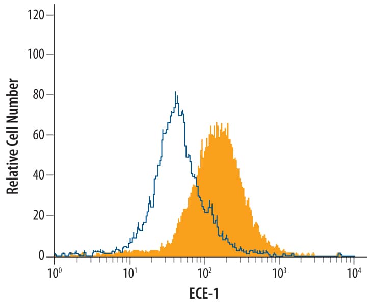
Detection of ECE-1 in MCF-7 Human Cell Line by Flow Cytometry.
MCF-7 human breast cancer cell line was stained with Human ECE-1 Antigen Affinity-purified Polyclonal Antibody (Catalog # AF1784, filled histogram) or control antibody (Catalog # AB-108-C, open histogram), followed by Allophycocyanin-conjugated Anti-Goat IgG Secondary Antibody (Catalog # F0108). To facilitate intracellular staining, cells were fixed with paraformaldehyde and permeabilized with saponin.ECE-1 in Human Kidney.
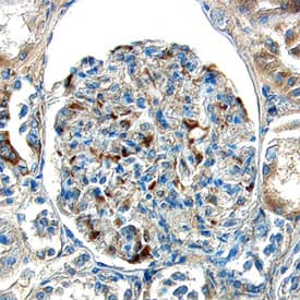
ECE-1 was detected in immersion fixed paraffin-embedded sections of human kidney using Goat Anti-Human ECE-1 Antigen Affinity-purified Polyclonal Antibody (Catalog # AF1784) at 15 µg/mL overnight at 4 °C. Tissue was stained using the Anti-Goat HRP-DAB Cell & Tissue Staining Kit (brown; Catalog # CTS008) and counterstained with hematoxylin (blue). Specific labeling was localized to the endothelial cells in glomeruli. View our protocol for Chromogenic IHC Staining of Paraffin-embedded Tissue Sections.Applications for Human ECE-1 Antibody
CyTOF-ready
Immunohistochemistry
Sample: Immersion fixed paraffin-embedded sections of human kidney
Immunoprecipitation
Sample: Conditioned cell culture medium spiked with Recombinant Human ECE‑1 (Catalog # 1784‑ZN), see our available Western blot detection antibodies
Intracellular Staining by Flow Cytometry
Sample: MCF-7 human breast cancer cell line fixed with paraformaldehyde and permeabilized with saponin
Western Blot
Sample: Recombinant Human ECE-1 (Catalog # 1784-ZN)
Formulation, Preparation, and Storage
Purification
Reconstitution
Formulation
Shipping
Stability & Storage
- 12 months from date of receipt, -20 to -70 °C as supplied.
- 1 month, 2 to 8 °C under sterile conditions after reconstitution.
- 6 months, -20 to -70 °C under sterile conditions after reconstitution.
Background: ECE-1
Endothelin-converting Enzyme 1 (ECE-1) is a zinc protease of the neprilysin (NEP) family, which also includes ECE-2, PEX, XCE, DINE, Kell and several NEP-like proteins (1). ECE-1 is a type II transmembrane protein with a short cytoplasmic tail and a large ectodomain. Four alternatively spliced isoforms differ in their cytoplasmic tail (2, 3). In addition to big endothelin-1, ECE-1 cleaves a variety of bioactive peptides such as bradykinin, neurotensin, angiotensin I, and substance P (1). Together with ECE-2, it is also involved in degradation of beta-amyloid peptide (4). The ectodomain of human ECE-1, which is common to all isoforms, was expressed with an N-terminal His tag and purified.
References
- Turner, A.J. et al. (2001) BioEssays 23:261.
- Valdennaire, O. et al. (1999) Eur. J. Biochem. 264:341.
- Schweizer, A. et al. (1997) Biochem. J. 328:871.
- Eckman, E.A. et al. (2003) J. Biol. Chem. 278:2081.
Long Name
Alternate Names
Entrez Gene IDs
Gene Symbol
UniProt
Additional ECE-1 Products
Product Documents for Human ECE-1 Antibody
Product Specific Notices for Human ECE-1 Antibody
For research use only
