Human EGFR PE-conjugated Antibody
R&D Systems, part of Bio-Techne | Catalog # FAB10951P


Key Product Details
Species Reactivity
Validated:
Cited:
Applications
Validated:
Cited:
Label
Antibody Source
Product Specifications
Immunogen
Leu25-Ser645
Accession # CAA25240
Specificity
Clonality
Host
Isotype
Scientific Data Images for Human EGFR PE-conjugated Antibody
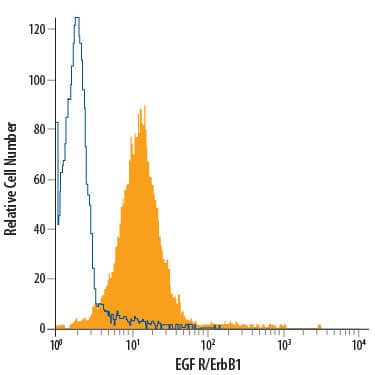
Detection of EGF R/ErbB1 in A431 human epithelial carcinoma cell line by Flow Cytometry.
A431 human epithelial carcinoma cell line was stained with Rat Anti-Human EGF R/ErbB1 PE-conjugated Monoclonal Antibody (Catalog # FAB10951P, filled histogram) or isotype control antibody (Catalog # IC006P, open histogram). View our protocol for Staining Membrane-associated Proteins.Applications for Human EGFR PE-conjugated Antibody
Flow Cytometry
Sample: A431 human epithelial carcinoma cell line
Formulation, Preparation, and Storage
Purification
Formulation
Shipping
Stability & Storage
- 12 months from date of receipt, 2 to 8 °C as supplied.
Background: EGFR
Epidermal Growth Factor Receptor (EGF R), also named erythroblastic leukemia viral oncogene homolog 1 (ErbB1), is a member of the type I receptor tyrosine kinase superfamily. The epidermal growth factor receptor (EGF R) subfamily of receptor tyrosine kinases comprises four members: EGF R (also known as HER1, ErbB1or ErbB), ErbB2 (Neu, HER2), ErbB3 (HER3), and ErbB4 (HER4). All family members are type I transmembrane glycoproteins that have an extracellular domain with two ligand binding cysteine rich domains, separated by a spacer region, and a cytoplasmic domain with a membrane proximal tyrosine kinase domain and a C-terminal tail with multiple tyrosine autophosphorylation sites. The human EGF R geneencodes a 1210 amino acid (aa) residue precursor with a 24 aa putative signal peptide, a 621 aa extracellular domain, a 23 aa transmembrane domain, and a 542 aa cytoplasmic domain. EGF R has been shown to bind a subset of the EGF family ligands, including EGF, amphiregulin, TGF alpha, betacellulin, epiregulin, heparin-binding EGF and neuregulin-2 alpha, in the absence of a coreceptor. Ligand binding induces EGF R homodimerization as well as heterodimerization with ErbB2, resulting in kinase activation, tyrosine phosphorylation and cell signaling. EGF R can also be recruited to form heterodimers with ligand-activated ErbB3 or ErbB4. EGF R signaling has been shown to regulate multiple biological functions including cell proliferation, differentiation, motility and apoptosis. In addition, EGF R signaling has also been shown to play a role in carcinogenesis (1 ‑ 3).
References
- Daly, R.J. (1999) Growth Factors, 16:255.
- Schlessinger, J. (2000) Cell. 103:211.
- Maihle, N.J. et al. (2002) Cancer Treat. Res. 107:247.
Long Name
Alternate Names
Gene Symbol
UniProt
Additional EGFR Products
Product Documents for Human EGFR PE-conjugated Antibody
Product Specific Notices for Human EGFR PE-conjugated Antibody
For research use only