Human EphA2 Antibody
R&D Systems, part of Bio-Techne | Catalog # AF3035


Key Product Details
Validated by
Species Reactivity
Validated:
Cited:
Applications
Validated:
Cited:
Label
Antibody Source
Product Specifications
Immunogen
Gln25-Asn534
Accession # P29317
Specificity
Clonality
Host
Isotype
Scientific Data Images for Human EphA2 Antibody
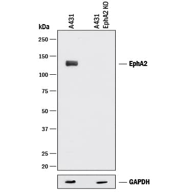
Detection of Human EphA2 by Western Blot.
Western blot shows lysates of A431 human epithelial carcinoma parental cell line and EphA2 knock out (KO) A431 cell line. PVDF membrane was probed with 1 µg/mL of Goat Anti-Human EphA2 Antigen Affinity-purified Polyclonal Antibody (Catalog # AF3035) followed by HRP-conjugated Anti-Goat IgG Secondary Antibody (HAF017). A specific band was detected for EphA2 at approximately 110 kDa (as indicated), but not detectable in the knockout A431 cell line. GAPDH (Catalog # MAB5718) is shown as a loading control. This experiment was conducted under reducing conditions and using Immunoblot Buffer Group 1.Detection of EphA2 in A431 Human Cell Line by Flow Cytometry.
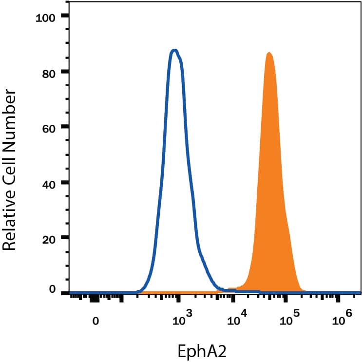
A431 human epithelial carcinoma cell line was stained with Goat Anti-Human EphA2 Antigen Affinity-purified Polyclonal Antibody (Catalog # AF3035, filled histogram) or isotype control antibody (Catalog # AB-108-C, open histogram), followed by Phycoerythrin-conjugated Anti-Goat IgG Secondary Antibody (Catalog # F0107). View our protocol for Staining Membrane-associated Proteins.EphA2 in Human Ovarian Cancer Tissue.
EphA2 was detected in immersion fixed paraffin-embedded sections of human ovarian cancer tissue using Goat Anti-Human EphA2 Antigen Affinity-purified Polyclonal Antibody (Catalog # AF3035) at 5 µg/mL overnight at 4 °C. Tissue was stained using the Anti-Goat HRP-DAB Cell & Tissue Staining Kit (brown; Catalog # CTS008) and counterstained with hematoxylin (blue). Specific labeling was localized to the plasma membrane of cancer cells. View our protocol for Chromogenic IHC Staining of Paraffin-embedded Tissue Sections.Applications for Human EphA2 Antibody
CyTOF-ready
Flow Cytometry
Sample: A431 human epithelial carcinoma cell line
Immunohistochemistry
Sample: Immersion fixed paraffin-embedded sections of human breast, ovarian, and pancreatic cancer tissue
Knockout Validated
Sample: A431 human epithelial carcinoma parental cell line and EphA2 knock out A431 cell line
Western Blot
Sample: A431 human epithelial carcinoma cell line
Reviewed Applications
Read 1 review rated 4 using AF3035 in the following applications:
Formulation, Preparation, and Storage
Purification
Reconstitution
Formulation
Shipping
Stability & Storage
- 12 months from date of receipt, -20 to -70 °C as supplied.
- 1 month, 2 to 8 °C under sterile conditions after reconstitution.
- 6 months, -20 to -70 °C under sterile conditions after reconstitution.
Background: EphA2
EphA2, also known as Eck, Myk2, and Sek2, is a member of the Eph receptor tyrosine kinase family which binds Ephrins A1, 2, 3, 4, and 5 (1-4). A and B class Eph proteins have a common structural organization. The human EphA2 cDNA encodes a 976 amino acid (aa) precursor including a 24 aa signal sequence, a 510 aa extracellular domain (ECD), a 24 aa transmembrane segment, and a 418 aa cytoplasmic domain. The ECD contains an N-terminal globular domain, a cysteine-rich domain, and two fibronectin type III domains (5). The cytoplasmic domain contains a juxtamembrane motif with two tyrosine residues, which are the major autophosphorylation sites, a kinase domain, and a sterile alpha motif (SAM) (5). The ECD of human EphA2 shares 90-94% aa sequence identity with mouse, bovine, and canine EphA2, and approximately 45% aa sequence identity with human EphA1, 3, 4, 5, 7, and 8. EphA2 becomes autophosphorylated following ligand binding (6, 7) and then interacts with SH2 domain-containing PI3-kinase to activate MAPK pathways (8, 9). Reverse signaling is also propagated through the Ephrin ligand. Transcription of EphA2 is dependent on the expression of E-Cadherin (10), and can be induced by p53 family transcription factors (11). EphA2 is upregulated in breast, prostate, and colon cancer vascular endothelium. Its ligand, EphrinA1, is expressed by the local tumor cells (12, 13). In some cases, EphA2 and EphrinA1 are expressed on the same blood vessels (14). EphA2 signaling cooperates with VEGF receptor signaling in promoting endothelial cell migration (13). The gene encoding human EphA2 maps to a region on chromosome 1 which is frequently deleted in neuroectodermal tumors (15).
References
- Poliakov, A. et al. (2004) Dev. Cell 7:465.
- Surawska, H. et al. (2004) Cytokine Growth Factor Rev. 15:419.
- Pasquale, E.B. (2005) Nat. Rev. Mol. Cell Biol. 6:462.
- Davy, A. and P. Soriano (2005) Dev. Dyn. 232:1.
- Bohme, B et al. (1993) Oncogene 8:2857.
- Pandey, A. et al. (1995) Science 268:567.
- Bartley, T.D. et al. (1994) Nature 368:558.
- Pandey, A. et al. (1994) J. Biol. Chem. 269:30154.
- Miao, H. et al. (2001) Nat. Cell Biol. 3:527.
- Orsulic, S. and R. Kemler (2000) J. Cell Sci. 113:1793.
- Dohn, M. et al. (2001) Oncogene 20:6503.
- Zelinski, D.P. et al. (2001) Cancer Res. 61:2301.
- Brantley, D.M. et al. (2002) Oncogene 21:7011.
- Ogawa, K. et al. (2000) Oncogene 19:6043.
- Sulman, E.P. et al. (1997) Genomics 40:371.
Alternate Names
Gene Symbol
UniProt
Additional EphA2 Products
Product Documents for Human EphA2 Antibody
Product Specific Notices for Human EphA2 Antibody
For research use only

