Human Erythropoietin/EPO Antibody
R&D Systems, part of Bio-Techne | Catalog # MAB2873

Key Product Details
Species Reactivity
Applications
Label
Antibody Source
Product Specifications
Immunogen
Ala28-Arg193
Accession # CAA26094
Specificity
Clonality
Host
Isotype
Scientific Data Images for Human Erythropoietin/EPO Antibody
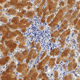
Erythropoietin/EPO in Human Liver.
Erythropoietin/EPO was detected in immersion fixed paraffin-embedded sections of human liver using Mouse Anti-Human Erythropoietin/EPO Monoclonal Antibody (Catalog # MAB2873) at 5 µg/mL for 1 hour at room temperature followed by incubation with the Anti-Mouse IgG VisUCyte™ HRP Polymer Antibody (Catalog # VC001). Before incubation with the primary antibody, tissue was subjected to heat-induced epitope retrieval using Antigen Retrieval Reagent-Basic (Catalog # CTS013). Tissue was stained using DAB (brown) and counterstained with hematoxylin (blue). Specific staining was localized to cytoplasm in hepatocytes. View our protocol for IHC Staining with VisUCyte HRP Polymer Detection Reagents.Applications for Human Erythropoietin/EPO Antibody
Immunohistochemistry
Sample: Immersion fixed paraffin-embedded sections of human liver
Formulation, Preparation, and Storage
Purification
Reconstitution
Formulation
Shipping
Stability & Storage
- 12 months from date of receipt, -20 to -70 °C as supplied.
- 1 month, 2 to 8 °C under sterile conditions after reconstitution.
- 6 months, -20 to -70 °C under sterile conditions after reconstitution.
Background: Erythropoietin/EPO
Erythropoietin (EPO) is a 34 kDa glycoprotein hormone in the type I cytokine family and is related to thrombopoietin (1). Its three N-glycosylation sites, four alpha helices, and N- to C-terminal disulfide bond are conserved across species (2, 3). Glycosylation of EPO is required for biological activities in vivo (4). Mature human EPO shares 75%-84% amino acid sequence identity with bovine, canine, equine, feline, mouse, ovine, porcine, and rat EPO. EPO is primarily produced in the kidney by a population of fibroblast-like cortical interstitial cells adjacent to the proximal tubules (5). It is also produced in much lower, but functionally significant amounts by fetal hepatocytes and in adult liver and brain (6-8). EPO promotes erythrocyte formation by preventing the apoptosis of early erythroid precursors which express the EPO receptor (EPO R) (8, 9). EPO R has also been described in brain, retina, heart, skeletal muscle, kidney, endothelial cells, and a variety of tumor cells (7, 8, 10, 11). Ligand induced dimerization of EPO R triggers JAK2-mediated signaling pathways followed by receptor/ligand endocytosis and degradation (1, 12). Rapid regulation of circulating EPO allows tight control of erythrocyte production and hemoglobin concentrations. Anemia or other causes of low tissue oxygen tension induce EPO production by stabilizing the hypoxia-induceable transcription factors HIF-1 alpha and HIF-2 alpha (1, 6). EPO additionally plays a tissue-protective role in ischemia by blocking apoptosis and inducing angiogenesis (7, 8, 13).
References
- Koury, M.J. (2005) Exp. Hematol. 33:1263.
- Jacobs, K. et al. (1985) Nature 313:806.
- Wen, D. et al. (1993) Blood 82:1507.
- Tsuda E., et al. (1990) Eur. J. Biochem. 188:405.
- Lacombe, C. et al. (1988) J. Clin. Invest. 81:620.
- Eckardt, K.U. and A. Kurtz (2005) Eur. J. Clin. Invest. 35 Suppl. 3:13.
- Sharples, E.J. et al. (2006) Curr. Opin. Pharmacol. 6:184.
- Rossert, J. and K. Eckardt (2005) Nephrol. Dial. Transplant 20:1025.
- Koury, M.J. and M.C. Bondurant (1990) Science 248:378.
- Acs, G. et al. (2001) Cancer Res. 61:3561.
- Hardee, M.E. et al. (2006) Clin. Cancer Res. 12:332.
- Verdier, F. et al. (2000) J. Biol. Chem. 275:18375.
- Kertesz, N. et al. (2004) Dev. Biol. 276:101.
Alternate Names
Gene Symbol
UniProt
Additional Erythropoietin/EPO Products
Product Documents for Human Erythropoietin/EPO Antibody
Product Specific Notices for Human Erythropoietin/EPO Antibody
For research use only
