Human FGFR3 Antibody
R&D Systems, part of Bio-Techne | Catalog # MAB766


Key Product Details
Validated by
Species Reactivity
Validated:
Cited:
Applications
Validated:
Cited:
Label
Antibody Source
Product Specifications
Immunogen
Specificity
Clonality
Host
Isotype
Scientific Data Images for Human FGFR3 Antibody
Detection of Mouse FGFR3 by Western Blot
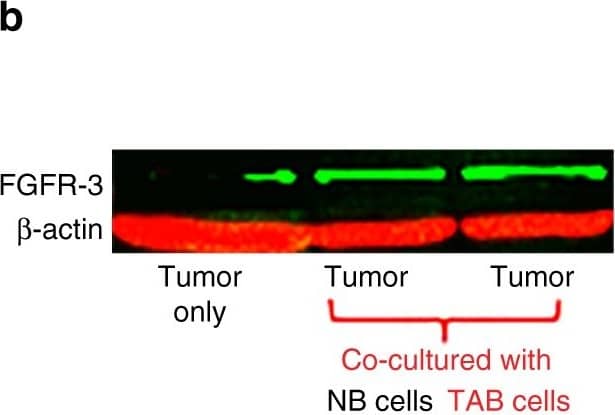
TAB cells modulate melanoma cells to express FGFR-3 and its ligand FGF-2 for tumor stroma-tumor cell cross-talk. a WM3749 co-cultured (72 h) with TAB cells (red bar) show increased FGFR-3 mRNA expression when compared with tumors only (open bar) or co-cultured with NB cells (blue bar). b Lysates obtained from pools of melanomas co-cultured (72–120 h) with NB- or TAB cells were probed in western blot with anti-FGFR-3 antibody (left panel), results expressed as relative intensity after beta-actin normalization (right panel). c Melanoma cells co-cultured with TAB cells (72 h) show increased phospho-FGFR-3 expression (right panel; immunofluorescence assays) when compared with melanoma cells alone (left panel) or melanoma cells co-cultured with NB cells (middle panel), scale bars: 40 μm, images captured by Nikon inverted microscope. d Melanoma cells co-cultured with TAB cells (red bar) show increased FGF-2 mRNA expression when compared with melanoma cells alone (open bar) or melanoma cells co-cultured with NB cells (blue bar). e 451Lu and WM989treated with IGF-1 (25 ng/ml/daily for 5 days; red bar) show increased FGFR-3 expression when compared with untreated controls (blue bar), flow cytometry results expressed as net % expression of control antibody. IGF-1 treated melanoma cells (red bars) show higher expression of FGFR-3 compared with untreated cells (blue bars). Bar represents mean + SD of replicate samples. f NB cells treated with FGF-2 (10 ng/ml/daily for 4 days; red bar) show high IGF-1 mRNA expression relative to untreated NB cells (blue bar). g 451Lu and WM989 co-cultured (72 h) with TAB cells in the presence of an anti-IGF-1 neutralizing antibody (10 μg/ml) show decreased FGFR-3 mRNA expression in tumor cells (blue bar) when compared with controls (red bar). h TAB cells co-cultured (72 h) with 451Lu and WM989 in the presence of an anti-FGF-2 neutralizing antibody (1 μg/ml) show decreased IGF-1 mRNA expression in B cells (blue bar) when compared with controls (red bar).Experiments in a, d and f–h were performed using qPCR. In Figures a, d–h, bars represent mean + SE of duplicate samples and are representative of at least two independent experiments. i Summary of cross-talk between melanoma and B cells: FGF-2 is constitutively expressed by tumor cells, released into the microenvironment to bind FGFR-3 on the B cells, activated B cells express increased levels of pro-inflammatory cytokines. IGF-1 released by TAB cells modulates tumor cells to increase their growth, heterogeneity and therapy resistance Image collected and cropped by CiteAb from the following publication (https://pubmed.ncbi.nlm.nih.gov/28928360), licensed under a CC-BY license. Not internally tested by R&D Systems.Applications for Human FGFR3 Antibody
CyTOF-ready
Flow Cytometry
Sample: K562 human chronic myelogenous leukemia cell line
Western Blot
Sample: Recombinant Human FGF R3 (IIIb) Fc Chimera (Catalog # 1264-FR)
Recombinant Human FGF R3 (IIIc) Fc Chimera (Catalog # 766-FR)
Reviewed Applications
Read 2 reviews rated 4 using MAB766 in the following applications:
Formulation, Preparation, and Storage
Purification
Reconstitution
Formulation
Shipping
Stability & Storage
- 12 months from date of receipt, -20 to -70 °C as supplied.
- 1 month, 2 to 8 °C under sterile conditions after reconstitution.
- 6 months, -20 to -70 °C under sterile conditions after reconstitution.
Background: FGFR3
Fibroblast Growth Factor Receptor 3 (FGF R3) is a type I transmembrane tyrosine kinase receptor that binds FGF ligands along with heparin or heparin sulfate proteoglycans as co‑factors. A segment of the membrane proximal Ig-like domain can be encoded by two different exons resulting in (IIIb) or (IIIc) isoforms. The IIIb or IIIc isoforms recognize FGF -1, -2, -4, -8b, -8e, -8f, -9, and -17b. FGF R3 plays a role in skeletal, brain, lung, intestine, kidney, and skin development.
Long Name
Alternate Names
Gene Symbol
Additional FGFR3 Products
Product Documents for Human FGFR3 Antibody
Product Specific Notices for Human FGFR3 Antibody
For research use only