Human Fibroblast Activation Protein alpha/FAP APC-conjugated Antibody
R&D Systems, part of Bio-Techne | Catalog # FAB3715A


Key Product Details
Species Reactivity
Validated:
Cited:
Applications
Validated:
Cited:
Label
Antibody Source
Product Specifications
Immunogen
Leu26-Asp760
Accession # Q12884
Specificity
Clonality
Host
Isotype
Scientific Data Images
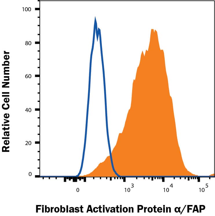
Detection of Fibroblast Activation Protein alpha/FAP in WI-38 Human Cell Line by Flow Cytometry.
WI-38 human lung fibroblast cell line was stained with Mouse Anti-Human Fibroblast Activation Protein a/FAP APC-conjugated Monoclonal Antibody (Catalog # FAB3715A, filled histogram) or isotype control antibody (IC002A, open histogram). View our protocol for Staining Membrane-associated Proteins.Detection of Fibroblast Activation Protein alpha/FAP in U87MG cells by Flow Cytometry.
U87MG cells were stained with Mouse Anti-Human Fibroblast Activation Protein alpha/FAP APC-conjugated Monoclonal Antibody (Catalog # FAB3715A, filled histogram) or isotype control antibody (Catalog # IC002A, open histogram). View our protocol for Staining Membrane-associated Proteins.Applications
Flow Cytometry
Sample: WI‑38 human lung fibroblast cell line and U87MG cells
Reviewed Applications
Read 1 review rated 5 using FAB3715A in the following applications:
Formulation, Preparation, and Storage
Purification
Formulation
Shipping
Stability & Storage
- 12 months from date of receipt, 2 to 8 °C as supplied.
Background: Fibroblast Activation Protein alpha/FAP
FAP (also known as Seprase) is a 97 kDa Type II transmembrane serine protease that is structurally related to Dipeptidyl Peptidase IV (DPPIV) (1). FAP has substrate specificity similar to DPPIV, which is specific for N-terminal Xaa-Pro sequences, but FAP is also an endopeptidase able to degrade gelatin and Type I Collagen (2). The enzymatically active form of FAP is a dimer that migrates at ~170 kDa. It is associated with multiple integral membrane proteins such as Integrin alpha3 beta1, UPA and DPPIV (3,4). FAP has a restricted tissue distribution. It is occasionally detected in fibroblasts and pancreatic islet cells, but is highly expressed on reactive stromal fibroblasts in epithelial cancers, in granulation tissue during wound healing, and in bone and soft tissue sarcomas (4-6). Because of its expression patterns and enzymatic activities, FAP is believed to play roles in tumor invasion, tissue remodeling, and wound repair. The 760 amino acid (aa) human FAP contains a 735 aa extracellular domain that is glycosylated and necessary for activity (4). It shares 90% aa identity with mouse and rat FAP. A reported 672 aa splicing variant diverges prior to the active site charge relay residues at the C-terminus.
References
- Scanlan, M.J. et al. (1994) Proc. Natl. Acad. Sci. USA 91:5657.
- Park, J.E. et al. (1999) J. Biol. Chem. 274:36505.
- Pineiro-Sanchez, M.L. et al. (1997) J. Biol. Chem. 272:7595.
- O'Brien, P. and B.F. O'Connor (2008) Biochim. Biophys. Acta 1784:1130.
- Garin-Chesa, P. et al. (1990) Proc. Natl. Acad. Sci. USA 87:7235.
- Rettig, W.J. et al. (1988) Proc. Natl. Acad. Sci. USA 85:3110.
Alternate Names
Gene Symbol
UniProt
Additional Fibroblast Activation Protein alpha/FAP Products
- All Products for Fibroblast Activation Protein alpha/FAP
- Fibroblast Activation Protein alpha/FAP cDNA Clones
- Fibroblast Activation Protein alpha/FAP ELISA Kits
- Fibroblast Activation Protein alpha/FAP Lysates
- Fibroblast Activation Protein alpha/FAP Primary Antibodies
- Fibroblast Activation Protein alpha/FAP Proteins and Enzymes
- Fibroblast Activation Protein alpha/FAP Simple Plex
Product Specific Notices
For research use only
