Human FOLR1 PE-conjugated Antibody
R&D Systems, part of Bio-Techne | Catalog # FAB5646P


Conjugate
Catalog #
Key Product Details
Validated by
Knockout/Knockdown
Species Reactivity
Validated:
Human
Cited:
Human
Applications
Validated:
Flow Cytometry, Knockout Validated
Cited:
Flow Cytometry, Immunocytochemistry
Label
Phycoerythrin (Excitation = 488 nm, Emission = 565-605 nm)
Antibody Source
Monoclonal Mouse IgG1 Clone # 548908
Product Specifications
Immunogen
Chinese hamster ovary cell line CHO-derived recombinant human FOLR1
Arg25-Met233
Accession # P15328
Arg25-Met233
Accession # P15328
Specificity
Detects human FOLR1 in direct ELISAs and Western blots. In direct ELISAs, no cross-reactivity with recombinant human FOLR2, 3 or 4 is observed.
Clonality
Monoclonal
Host
Mouse
Isotype
IgG1
Scientific Data Images for Human FOLR1 PE-conjugated Antibody
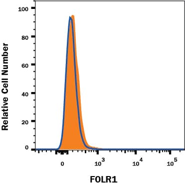
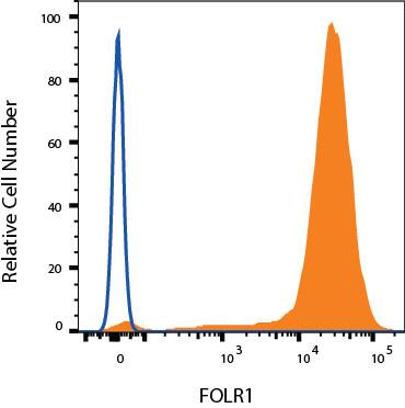
Detection of FOLR1 in MDA-MB-231 Human Cell Line by Flow Cytometry.
MDA-MB-231 human breast cancer cell line was stained with Mouse Anti-Human FOLR1 PE-conjugated Monoclonal Antibody (Catalog # FAB5646P, filled histogram) or isotype control antibody (IC002P, open histogram). View our protocol for Staining Membrane-associated Proteins.Detection of FOLR1 in MCF-7 Human Cell Line by Flow Cytometry.
MCF-7 human breast cancer cell line was stained with Mouse Anti-Human FOLR1 PE-conjugated Monoclonal Antibody (Catalog # FAB5646P, filled histogram) or isotype control antibody (IC002P, open histogram). View our protocol for Staining Membrane-associated Proteins.FOLR1 Specificity is Shown by Flow Cytometry in Knockout Cell Line.
FOLR1 knockout MCF-7 human breast cancer cell line was stained with Mouse Anti-Human FOLR1 PE-conjugated Monoclonal Antibody (Catalog # FAB5646P, filled histogram) or isotype control antibody (IC002P, open histogram). No staining in the FOLR1 knockout MCF-7 cell line was observed. View our protocol for Staining Membrane-associated Proteins.Applications for Human FOLR1 PE-conjugated Antibody
Application
Recommended Usage
Flow Cytometry
10 µL/106 cells
Sample: MDA‑MB‑231 and MCF-7 human breast cancer cell lines; HeLa human cervical epithelial carcinoma cell line
Sample: MDA‑MB‑231 and MCF-7 human breast cancer cell lines; HeLa human cervical epithelial carcinoma cell line
Knockout Validated
FOLR1 is specifically detected in MCF-7 human breast cancer parental cell line but is not detectable in FOLR1 knockout MCF-7 cell line.
Reviewed Applications
Read 1 review rated 4 using FAB5646P in the following applications:
Formulation, Preparation, and Storage
Purification
Protein A or G purified from hybridoma culture supernatant
Formulation
Supplied in a saline solution containing BSA and Sodium Azide.
Shipping
The product is shipped with polar packs. Upon receipt, store it immediately at the temperature recommended below.
Stability & Storage
Protect from light. Do not freeze.
- 12 months from date of receipt, 2 to 8 °C as supplied.
Background: FOLR1
References
- Kelemen, L.E. (2006) Int. J. Cancer 119:243.
- Fowler, B. (2001) Kidney Int. Suppl. 78:S221.
- Luhrs, C.A. et al. (1989) J. Biol. Chem. 264:21446.
- Lacey, S.W. et al. (1989) J. Clin. Invest. 84:715.
- Elwood, P.C. (1989) J. Biol. Chem. 264:14893.
- Rijnboutt, S. et al. (1996) J. Cell Biol. 132:35.
- Ross, J.F. et al. (1994) Cancer 73:2432.
- Parker, N. et al. (2005) Anal. Biochem. 338:284.
- Piedrahita, J.A. et al. (1999) Nat. Genet. 23:228.
- Paulos, C.M. et al. (2004) Mol. Pharmacol. 66:1406.
- Elwood, P.C. et al. (1991) J. Biol. Chem. 26:2346.
Long Name
Folate Receptor 1
Alternate Names
FBP, Folbp1, FR-alpha, MOv18
Gene Symbol
FOLR1
UniProt
Additional FOLR1 Products
Product Documents for Human FOLR1 PE-conjugated Antibody
Product Specific Notices for Human FOLR1 PE-conjugated Antibody
For research use only
Loading...
Loading...
Loading...
Loading...
Loading...


