Human FoxP1 Antibody
R&D Systems, part of Bio-Techne | Catalog # MAB45341

Key Product Details
Species Reactivity
Validated:
Cited:
Applications
Validated:
Cited:
Label
Antibody Source
Product Specifications
Immunogen
Lys548-Glu677
Accession # Q9H334
Specificity
Clonality
Host
Isotype
Scientific Data Images for Human FoxP1 Antibody
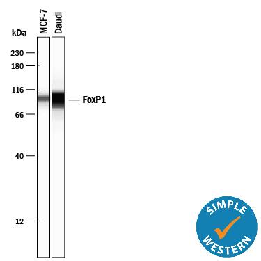
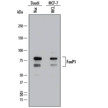
Detection of Human FoxP1 by Western Blot.
Western blot shows lysates of Daudi human Burkitt's lymphoma cell line and MCF-7 human breast cancer cell line. Gels were loaded with 25 µg of whole cell lysate (WCL) and 25 µg of nuclear extracts (Nuc). PVDF membrane was probed with 1 µg/mL of Mouse Anti-Human FoxP1 Monoclonal Antibody (Catalog # MAB45341) followed by HRP-conjugated Anti-Mouse IgG Secondary Antibody (Catalog # HAF018). Specific bands were detected for FoxP1 at approximately 65 and 80 kDa (as indicated). This experiment was conducted under reducing conditions and using Immunoblot Buffer Group 1.FoxP1 in BG01V Human Embryonic Stem Cells.
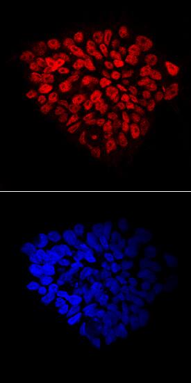
FoxP1 was detected in immersion fixed BG01V human embryonic stem cells using Mouse Anti-Human FoxP1 Monoclonal Antibody (Catalog # MAB45341) at 10 µg/mL for 3 hours at room temperature. Cells were stained using the NorthernLights™ 557-conjugated Anti-Mouse IgG Secondary Antibody (red, upper panel; Catalog # NL007) and counterstained with DAPI (blue, lower panel). Specific staining was localized to nuclei. View our protocol for Fluorescent ICC Staining of Stem Cells on Coverslips.FoxP1 in Human Prostate.
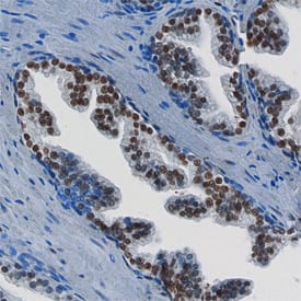
FoxP1 was detected in immersion fixed paraffin-embedded sections of human prostate using Mouse Anti-Human FoxP1 Monoclonal Antibody (Catalog # MAB45341) at 15 µg/mL overnight at 4 °C. Before incubation with the primary antibody, tissue was subjected to heat-induced epitope retrieval using Antigen Retrieval Reagent-Basic (Catalog # CTS013). Tissue was stained using the Anti-Mouse HRP-DAB Cell & Tissue Staining Kit (brown; Catalog # CTS002) and counter-stained with hematoxylin (blue). Specific staining was localized to the nuclei of epithelial cells. View our protocol for Chromogenic IHC Staining of Paraffin-embedded Tissue Sections.Applications for Human FoxP1 Antibody
CyTOF-ready
Immunocytochemistry
Sample: Immersion fixed BG01V human embryonic stem cells
Immunohistochemistry
Sample: Immersion fixed paraffin-embedded sections of human prostate subjected to heat-induced epitope retrieval using Antigen Retrieval Reagent-Basic (Catalog # CTS013)
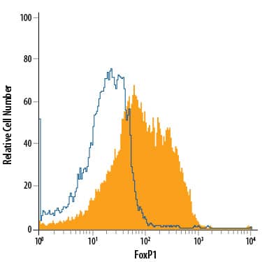
Intracellular Staining by Flow Cytometry
Sample: MCF‑7 human breast cancer cell line fixed with 4% paraformaldehyde and permeabilized with methanol
Simple Western
Sample: MCF‑7 human breast cancer cell line and Daudi human Burkitt's lymphoma cell line
Western Blot
Sample: Daudi human Burkitt's lymphoma cell line and MCF‑7 human breast cancer cell line
Formulation, Preparation, and Storage
Purification
Reconstitution
Formulation
Shipping
Stability & Storage
- 12 months from date of receipt, -20 to -70 °C as supplied.
- 1 month, 2 to 8 °C under sterile conditions after reconstitution.
- 6 months, -20 to -70 °C under sterile conditions after reconstitution.
Background: FoxP1
Forkhead Box P1 (FOXP1) is a member of the FOX family of transcription factors. FoxP1 has been implicated in cardiac, lung, and lymphocyte development. FoxP1 knock out mice die at embryonic day 14.5 due to heart valve and outflow tract abnormalities. FoxP1 contains both a DNA binding domain as well as protein-protein interaction domains. FoxP1 can homo or heterodimerize with FoxP2 and FoxP4, with dimerization necessary for DNA binding. FoxP1 shows both oncogenic and tumor suppressive characteristics. Overexpression in lymphomas leads to poor prognosis, but loss of FoxP1 in breast cancer also implicates a poor prognosis. Human isoforms of 489 to 677 amino acids contain alternate sequences within the first 60 amino acids and/or deletion of amino acids.
Long Name
Alternate Names
Gene Symbol
UniProt
Additional FoxP1 Products
Product Documents for Human FoxP1 Antibody
Product Specific Notices for Human FoxP1 Antibody
For research use only
