Human Glut1 Fluorescein-conjugated Antibody
R&D Systems, part of Bio-Techne | Catalog # FAB1418F


Conjugate
Catalog #
Key Product Details
Validated by
Biological Validation
Species Reactivity
Validated:
Human
Cited:
Human
Applications
Validated:
Flow Cytometry
Cited:
Flow Cytometry
Label
Fluorescein (Excitation = 488 nm, Emission = 515-545 nm)
Antibody Source
Monoclonal Mouse IgG2B Clone # 202915
Product Specifications
Immunogen
NS0 mouse myeloma cell line transfected with human Glut1
Met1-Val492
Accession # AAA52571
Met1-Val492
Accession # AAA52571
Specificity
Detects human Glut1. Stains human Glut1-transfected NS0 cells, but not NS0 control transfectants. Although
Human Glut1 Antibody detects Glut1 on the surface of T cells (1, 2), it does not detect it on erythrocytes (5). The reason for this discrepancy is not understood, but may be related to conformational or post-translational modification differences.
Clonality
Monoclonal
Host
Mouse
Isotype
IgG2B
Scientific Data Images for Human Glut1 Fluorescein-conjugated Antibody
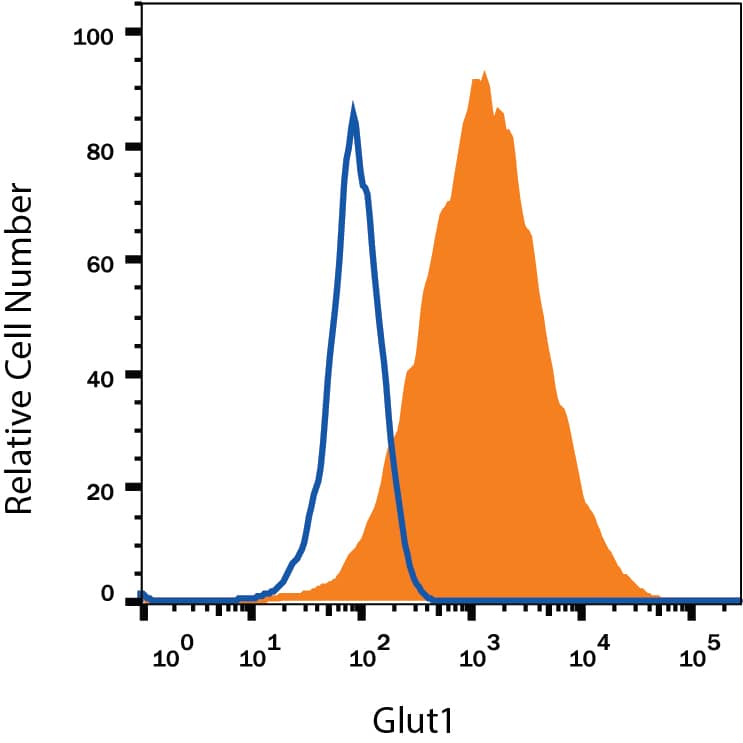
Detection of Glut1 in HepG2 Human Cell Line by Flow Cytometry.
HepG2 human hepatocellular carcinoma cell line was stained with Mouse Anti-Human Glut1 Fluorescein-conjugated Monoclonal Antibody (Catalog # FAB1418F, filled histogram) or isotype control antibody (Catalog # IC0041F, open histogram). View our protocol for Staining Membrane-associated Proteins.Detection of Glut1 in Jurkat Human Cell Line by Flow Cytometry.
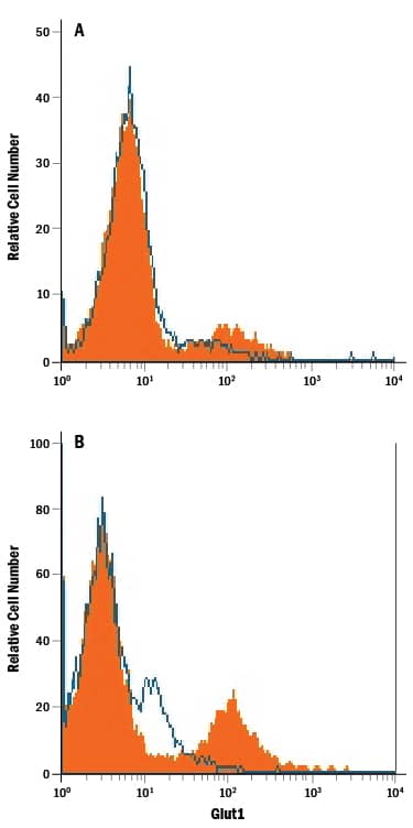
Jurkat human acute T cell leukemia cell line either (A) untreated or (B) cultured in nutrient-depleted media was stained with Mouse Anti-Human Glut1 Fluorescein-conjugated Monoclonal Antibody (Catalog # FAB1418F, filled histogram) or isotype control antibody (Catalog # IC0041F, open histogram). View our protocol for Staining Membrane-associated Proteins.Applications for Human Glut1 Fluorescein-conjugated Antibody
Application
Recommended Usage
Flow Cytometry
10 µL/106 cells
Sample: HepG2 human hepatocellular carcinoma cell line (untreated) and Jurkat human acute T cell leukemia cell line cultured in nutrient-depleted media
Sample: HepG2 human hepatocellular carcinoma cell line (untreated) and Jurkat human acute T cell leukemia cell line cultured in nutrient-depleted media
Formulation, Preparation, and Storage
Purification
Protein A or G purified from hybridoma culture supernatant
Formulation
Supplied in a saline solution containing BSA and Sodium Azide.
Shipping
The product is shipped with polar packs. Upon receipt, store it immediately at the temperature recommended below.
Stability & Storage
Protect from light. Do not freeze.
- 12 months from date of receipt, 2 to 8 °C as supplied.
Background: Glut1
Glut1 belongs to the facilitative glucose transport protein family that comprises 13 members. It is an integral membrane protein with 12 transmembrane domains and is expressed at variable levels in many tissues including brain endothelial cells, CD8+ T cells, and erythrocytes (1‑4). Glut1 is a major glucose transporter that mediates glucose transport across the mammalian blood‑brain barrier.
References
- Mueckler, M. et al. 1994, Eur. J. Biochem. 219:713.
- Meuckler, M. et al. 1985, Science 229:941.
- Jones, K.S. et al. 2006, J. Virol. 8291.
- Takenouchi, N. et al. 2007, J. Virol. 1506.
- Kinet, S. et al. 2007, Retrovirology 4:31.
Long Name
Glucose Transporter Type 1
Alternate Names
DYT17, DYT18, DYT9, EIG12, GLUT1DS, SLC2A1
Gene Symbol
SLC2A1
UniProt
Additional Glut1 Products
Product Documents for Human Glut1 Fluorescein-conjugated Antibody
Product Specific Notices for Human Glut1 Fluorescein-conjugated Antibody
For research use only
Loading...
Loading...
Loading...
Loading...
Loading...
