Human Hematopoietic Prostaglandin D Synthase/HPGDS Antibody
R&D Systems, part of Bio-Techne | Catalog # MAB6487

Key Product Details
Species Reactivity
Applications
Label
Antibody Source
Product Specifications
Immunogen
Pro2-Leu199
Accession # O60760
Specificity
Clonality
Host
Isotype
Scientific Data Images for Human Hematopoietic Prostaglandin D Synthase/HPGDS Antibody
Detection of Human Hematopoietic Prostaglandin D Synthase/HPGDS by Western Blot.
Western blot shows lysates of NCI-H460 human large cell lung carcinoma cell line, human lung tissue, and human placenta tissue. PVDF membrane was probed with 0.25 µg/mL of Mouse Anti-Human Hematopoietic Prostaglandin D Synthase/HPGDS Monoclonal Antibody (Catalog # MAB6487) followed by HRP-conjugated Anti-Mouse IgG Secondary Antibody (Catalog # HAF007). A specific band was detected for Hematopoietic Prostaglandin D Synthase/HPGDS at approximately 25 kDa (as indicated). This experiment was conducted under reducing conditions and using Immunoblot Buffer Group 1.Hematopoietic Prostaglandin D Synthase/HPGDS in HEL 92.1.7 Human Cell Line.
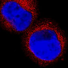
Hematopoietic Prostaglandin D Synthase/HPGDS was detected in immersion fixed HEL 92.1.7 human erythroleukemic cell line using Mouse Anti-Human Hematopoietic Prostaglandin D Synthase/HPGDS Monoclonal Antibody (Catalog # MAB6487) at 8 µg/mL for 3 hours at room temperature. Cells were stained using the NorthernLights™ 557-conjugated Anti-Mouse IgG Secondary Antibody (red; Catalog # NL007) and counterstained with DAPI (blue). Specific staining was localized to cytoplasm. View our protocol for Fluorescent ICC Staining of Non-adherent Cells.Applications for Human Hematopoietic Prostaglandin D Synthase/HPGDS Antibody
Immunocytochemistry
Sample: Immersion fixed HEL 92.1.7 human erythroleukemic cell line
Western Blot
Sample: NCI‑H460 human large cell lung carcinoma cell line, human lung tissue, and human placenta tissue
Reviewed Applications
Read 1 review rated 3 using MAB6487 in the following applications:
Formulation, Preparation, and Storage
Purification
Reconstitution
Formulation
Shipping
Stability & Storage
- 12 months from date of receipt, -20 to -70 °C as supplied.
- 1 month, 2 to 8 °C under sterile conditions after reconstitution.
- 6 months, -20 to -70 °C under sterile conditions after reconstitution.
Background: Hematopoietic Prostaglandin D Synthase/HPGDS
Prostaglandin D Synthase (PGDS) catalyzes the conversion of prostaglandin (PG) H2 to PGD2, which is a major prostanoid produced in a variety of tissues. Two types of PGDS have been isolated; the glutathione-dependent hematopoietic PGDS (HPGDS) and the glutathione-independent lipocalin‑type PGDS (1). HPGDS is a cytosolic enzyme that is expressed in mast cells and antigen presenting cells (2, 3). It is the only mammalian member of the class Sigma glutathione S‑transferase, showing a broad specificity towards standard transferase substrates (4). The PGD2 produced by HPGDS is involved in many physiological processes such as maintaining body temperature, promotion of sleep, inhibition of platelet aggregation and bronchoconstriction (5). It also functions in immune response and acts as a mediator in allergy and inflammation (6). HPGDS‑specific inhibitors may be therapeutically useful anti‑allergic and anti‑inflammatory drugs.
References
- Urade, Y. and Eguchi, N. (2002) Prostaglandins Other Lipid Mediat. 68:375.
- Urade, Y. et al. (1990) J. Biol. Chem. 265:371.
- Urade, Y. et al. (1989) J. Immunol. 143:2982.
- Jowsey, I. R. et al. (2001) Biochem. J. 359:507.
- Kanaoka, Y. and Urade, Y. (2003) Prostaglandins Leukot. Essent. Fatty Acids 69:163.
- Oguma, T. et al. (2008) Allergol. Int. 57:307.
Alternate Names
Gene Symbol
UniProt
Additional Hematopoietic Prostaglandin D Synthase/HPGDS Products
- All Products for Hematopoietic Prostaglandin D Synthase/HPGDS
- Hematopoietic Prostaglandin D Synthase/HPGDS ELISA Kits
- Hematopoietic Prostaglandin D Synthase/HPGDS Lysates
- Hematopoietic Prostaglandin D Synthase/HPGDS Primary Antibodies
- Hematopoietic Prostaglandin D Synthase/HPGDS Proteins and Enzymes
Product Documents for Human Hematopoietic Prostaglandin D Synthase/HPGDS Antibody
Product Specific Notices for Human Hematopoietic Prostaglandin D Synthase/HPGDS Antibody
For research use only

