Human IGF-II/IGF2 Antibody
R&D Systems, part of Bio-Techne | Catalog # AF-292-NA


Key Product Details
Species Reactivity
Validated:
Cited:
Applications
Validated:
Cited:
Label
Antibody Source
Product Specifications
Immunogen
Ala25-Glu91
Accession # P01344
Specificity
Clonality
Host
Isotype
Endotoxin Level
Scientific Data Images for Human IGF-II/IGF2 Antibody
Cell Proliferation Induced by IGF-II/IGF2 and Neutralization by Human IGF-II/IGF2 Antibody.
Recombinant Human IGF-II/IGF2 (Catalog # 292-G2) stimulates proliferation in the MCF-7 human breast cancer cell line in a dose-dependent manner (orange line). Proliferation elicited by Recombinant Human IGF-II/IGF2 (14 ng/mL) is neutralized (green line) by increasing concentrations of Goat Anti-Human IGF-II/IGF2 Antigen Affinity-purified Polyclonal Antibody (Catalog # AF-292-NA). The ND50 is typically 0.2-0.8 µg/mL.IGF-II/IGF2 in Human Placenta.
IGF-II/IGF2 was detected in immersion fixed paraffin-embedded sections of human placenta (chorionic villi) using 5 µg/mL Goat Anti-Human IGF-II/IGF2 Antigen Affinity-purified Polyclonal Antibody (Catalog # AF-292-NA) overnight at 4 °C. Before incubation with the primary antibody tissue was subjected to heat-induced epitope retrieval using Antigen Retrieval Reagent-Basic (Catalog # CTS013). Tissue was stained with the Anti-Goat HRP-AEC Cell & Tissue Staining Kit (red; Catalog # CTS009) and counterstained with hematoxylin (blue). View our protocol for Chromogenic IHC Staining of Paraffin-embedded Tissue Sections.Detection of Human IGF-II/IGF2 by Western Blot
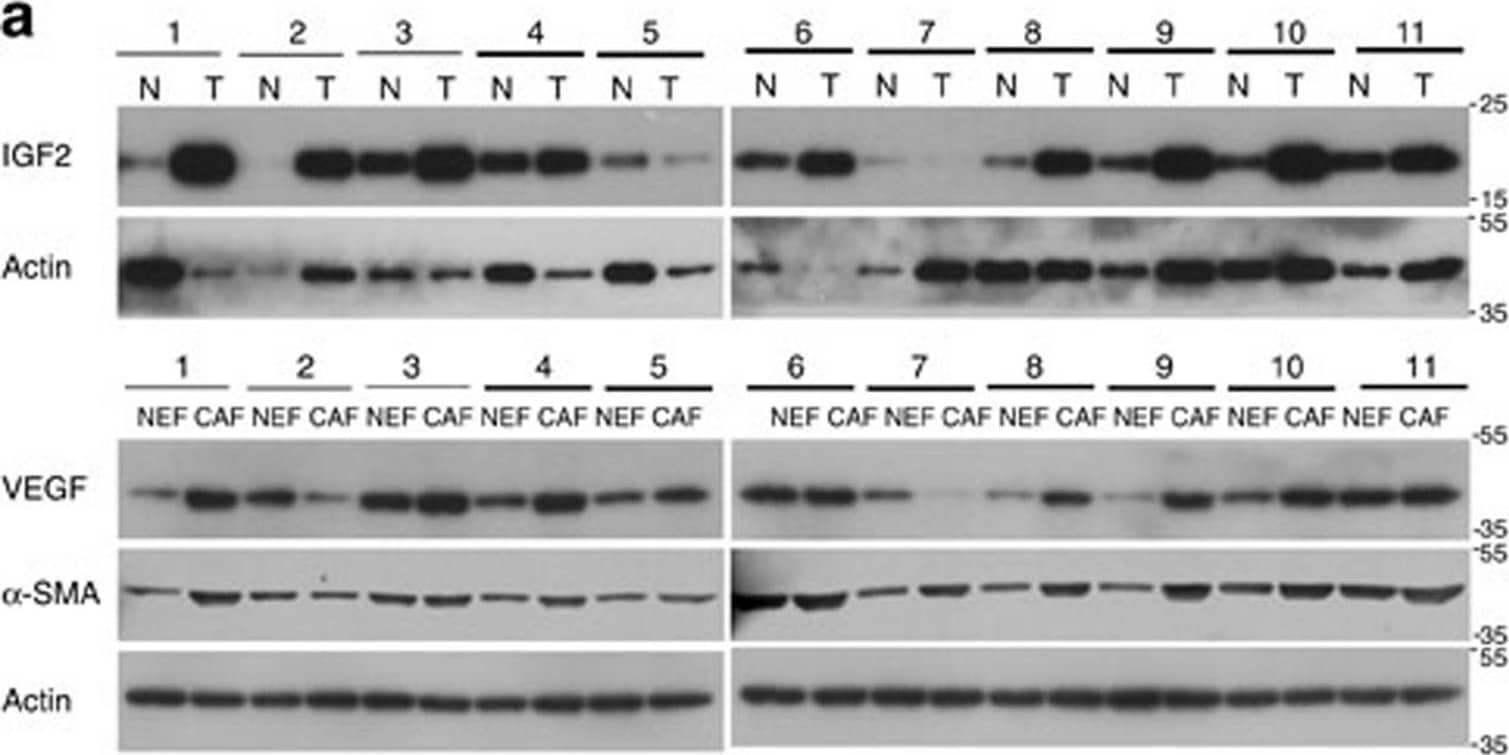
Clinical relevance of tumour IGF2 and stromal VEGF axis.(a) Western blot showing expression of IGF2 in ESCC (T) and matched non-tumour specimens (N), as well as expression of VEGF and alpha-SMA in CAFs and matched NEF from 11 ESCC patients. (b) Graphs showing positive correlation between IGF2 expression in oesophageal tissue and expressions of VEGF (left panel) and alpha-SMA (right panel) in fibroblasts, respectively, in 11 pairs of ESCC and adjacent normal tissues. Correlation was assessed using Pearson's rank correlation coefficient. (c) Comparison of serum IGF2 (left panel) and VEGF (middle panel) levels between healthy individuals (n=50) and ESCC patients (n=100); the data were pooled and a positive correlation was found between serum VEGF and IGF2 levels (n=150) (right panel) using unpaired t-test. (d) Gene expressions were further divided into high and low levels using median expression level as the cut-off point for survival analyses. Kaplan–Meier curves comparing survival outcome of ESCC patients (n=100) with high and low serum IGF2 levels (left panel), high and low serum VEGF levels (middle panel), and IGF2 High/VEGF High and IGF2 Low/VEGF Low levels (right panel); statistical significance was calculated by log-rank test. Image collected and cropped by CiteAb from the following open publication (https://www.nature.com/articles/ncomms14399), licensed under a CC-BY license. Not internally tested by R&D Systems.Applications for Human IGF-II/IGF2 Antibody
Immunohistochemistry
Sample: Immersion fixed paraffin-embedded sections of human placenta (chorionic villi) subjected to Antigen Retrieval Reagent-Basic (Catalog # CTS013)
Western Blot
Sample: Recombinant Human IGF-II/IGF2 (Catalog # 292-G2)
Neutralization
Formulation, Preparation, and Storage
Purification
Reconstitution
Formulation
Shipping
Stability & Storage
- 12 months from date of receipt, -20 to -70 °C as supplied.
- 1 month, 2 to 8 °C under sterile conditions after reconstitution.
- 6 months, -20 to -70 °C under sterile conditions after reconstitution.
Background: IGF-II/IGF2
Insulin-like growth factor I (also known as somatomedin C and somatomedin A) and insulin-like growth factor II (multiplication stimulating activity or MSA) belong to the family of insulin-like growth factors that are structurally homologous to proinsulin. Mature IGF-I and IGF-II share approximately 70% sequence identity. Both IGF-I and IGF-II are expressed in many tissues and cell types and may have autocrine, paracrine and endocrine functions. Mature IGF-I and IGF-II are highly conserved (100% identity between human, bovine, and porcine proteins) and exhibit cross-species activity.
IGF-II is a potent mitogenic growth factor. However, unlike IGF-I which has important postnatal roles, the growth-promoting function of IGF-II is limited to embryonic development.
Two specific cell surface receptors that bind IGF-I and IGF-II have been identified. The type I IGF receptor that participates in IGF signaling is structurally related to the insulin receptor. It is a disulfide-linked heterotetrameric transmembrane glycoprotein with an intracellular tyrosine kinase domain. Type I IGF receptor binds IGF-I with higher affinity than IGF-II. The type II IGF receptor which binds IGF-II with much higher affinity than IGF-I is also the cation-independent mannose 6-phosphate receptor. At the present time, it is not known if the type II IGF receptor participates in the IGF signaling pathway. An additional unknown receptor which mediates IGF-II signaling has also been proposed. Circulating IGFs exist in complexes bound to IGF binding proteins. Currently, at least six high affinity binding proteins have been identified.
Long Name
Alternate Names
Gene Symbol
UniProt
Additional IGF-II/IGF2 Products
Product Documents for Human IGF-II/IGF2 Antibody
Product Specific Notices for Human IGF-II/IGF2 Antibody
For research use only

