Human IGFBP-5 Antibody
R&D Systems, part of Bio-Techne | Catalog # AF875


Key Product Details
Species Reactivity
Validated:
Cited:
Applications
Validated:
Cited:
Label
Antibody Source
Product Specifications
Immunogen
Glu28-Glu272
Accession # P24593
Specificity
Clonality
Host
Isotype
Scientific Data Images for Human IGFBP-5 Antibody
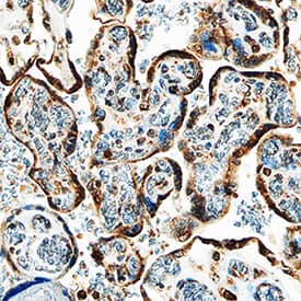
IGFBP‑5 in Human Placenta.
IGFBP-5 was detected in immersion fixed paraffin-embedded sections of human placenta using Goat Anti-Human IGFBP-5 Antigen Affinity-purified Polyclonal Antibody (Catalog # AF875) at 1.7 µg/mL overnight at 4 °C. Tissue was stained using the Anti-Goat HRP-DAB Cell & Tissue Staining Kit (brown; Catalog # CTS008) and counterstained with hematoxylin (blue). Specific staining was localized to syncytiotrophoblasts. View our protocol for Chromogenic IHC Staining of Paraffin-embedded Tissue Sections.Detection of Human IGFBP-5 by Immunohistochemistry
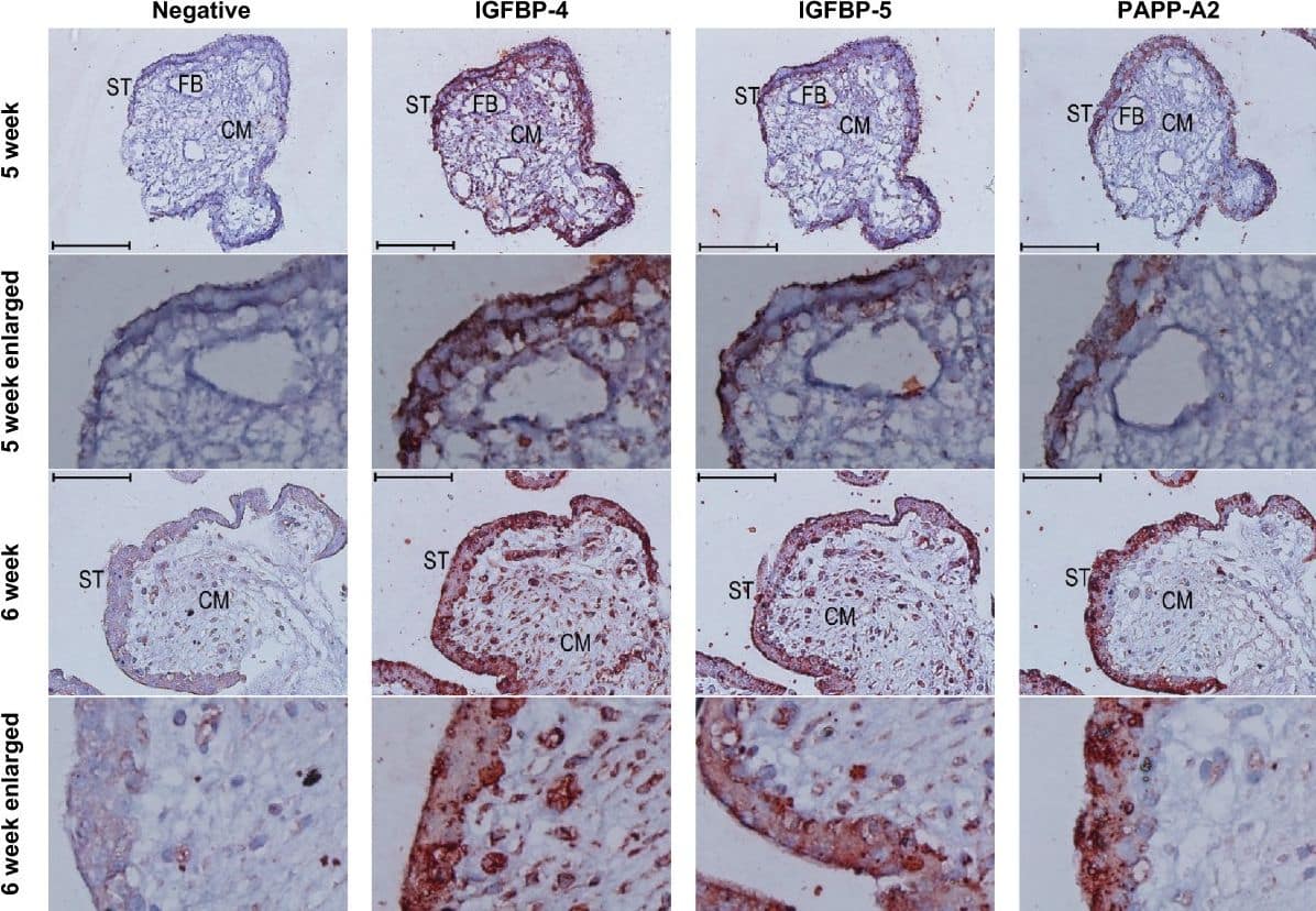
Immunoreactivity (red colour) against IGFBP-4, −5, and PAPPA2 in the syncytiotrophoblast of first trimester placental villi. Serial cross sections of placental villi at 5 and 6 weeks of gestation (7 and 13 weeks not shown) were stained for IGFBP-4, IGFBP-5, and PAPP-A2. Negative controls used non-specific goat IgG in the place of the primary antibodies. FB = fetal blood vessel, CM = chorionic mesoderm, ST = syncytiotrophoblast. Scale bars denote 100 μm. Image collected and cropped by CiteAb from the following open publication (https://pubmed.ncbi.nlm.nih.gov/25475528), licensed under a CC-BY license. Not internally tested by R&D Systems.Detection of Human IGFBP-5 by Immunohistochemistry
Immunoreactivity (red colour) against IGFBP-4, −5, and PAPPA2 in the syncytiotrophoblast of first trimester placental villi. Serial cross sections of placental villi at 5 and 6 weeks of gestation (7 and 13 weeks not shown) were stained for IGFBP-4, IGFBP-5, and PAPP-A2. Negative controls used non-specific goat IgG in the place of the primary antibodies. FB = fetal blood vessel, CM = chorionic mesoderm, ST = syncytiotrophoblast. Scale bars denote 100 μm. Image collected and cropped by CiteAb from the following open publication (https://pubmed.ncbi.nlm.nih.gov/25475528), licensed under a CC-BY license. Not internally tested by R&D Systems.Applications for Human IGFBP-5 Antibody
Immunohistochemistry
Sample: Immersion fixed paraffin-embedded sections of human placenta
Western Blot
Sample: Recombinant Human IGFBP-5 (Catalog # 875-B5)
Formulation, Preparation, and Storage
Purification
Reconstitution
Formulation
Shipping
Stability & Storage
- 12 months from date of receipt, -20 to -70 °C as supplied.
- 1 month, 2 to 8 °C under sterile conditions after reconstitution.
- 6 months, -20 to -70 °C under sterile conditions after reconstitution.
Background: IGFBP-5
The superfamily of insulin-like growth factor (IGF) binding proteins include the six high-affinity IGF binding proteins (IGFBP) and at least four additional low-affinity binding proteins referred to as IGFBP related proteins (IGFBP-rP). All IGFBP superfamily members are cysteine-rich proteins with conserved cysteine residues, which are clustered in the amino- and carboxy-terminal thirds of the molecule. IGFBPs modulate the biological activities of IGF proteins. Some IGFBPs may also have intrinsic bioactivity that is independent of their ability to bind IGF proteins. Post-transitional modifications of IGFBP, including glycosylation, phosphorylation and proteolysis, have been shown to modify the affinities of the binding proteins to IGF.
Human IGFBP-5 cDNA encodes a 272 amino acid (aa) residue precursor protein with a putative 20 aa residue signal peptide that is processed to generate the 252 aa residue mature protein that is post-translationally modified by O-glycosylations and serine phosphorylations. IGFBP-5 is expressed by fibroblasts, myoblasts and osteoblasts, making it the predominant IGFBP found in bone extracts. IGFBP-5 has a strong affinity for hydroxyapatite, allowing it to bind to bone cells. When bound to extracellular matrix, IGFBP-5 is protected from proteolysis and potentiates IGF activity, but when it is soluble, IGFBP-5 is cleaved to a biologically inactive 21 kDa fragment (1, 2).
References
- Jones, J.I. and D.R. Clemmons (1995) Endocrine Rev. 16:3.
- Kelley, K.M. et al. (1996) Int. J. Biochem. Cell Biol. 28:619.
Long Name
Alternate Names
Gene Symbol
UniProt
Additional IGFBP-5 Products
Product Documents for Human IGFBP-5 Antibody
Product Specific Notices for Human IGFBP-5 Antibody
For research use only

