Human IGFBP-rp1/IGFBP-7 Antibody
R&D Systems, part of Bio-Techne | Catalog # AF1334


Key Product Details
Species Reactivity
Validated:
Cited:
Applications
Validated:
Cited:
Label
Antibody Source
Product Specifications
Immunogen
Arg98-Arg277
Accession # AAA16187
Specificity
Clonality
Host
Isotype
Scientific Data Images for Human IGFBP-rp1/IGFBP-7 Antibody
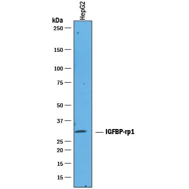
Detection of Human IGFBP‑rp1/IGFBP‑7 by Western Blot.
Western blot shows lysates of HepG2 human hepatocellular carcinoma cell line. PVDF membrane was probed with 1 µg/mL of Goat Anti-Human IGFBP-rp1/IGFBP-7 Antigen Affinity-purified Polyclonal Antibody (Catalog # AF1334) followed by HRP-conjugated Anti-Goat IgG Secondary Antibody (Catalog # HAF019). A specific band was detected for IGFBP-rp1/IGFBP-7 at approximately 30 kDa (as indicated). This experiment was conducted under reducing conditions and using Immunoblot Buffer Group 1.IGFBP‑rp1/IGFBP‑7 in Human Pancreas.
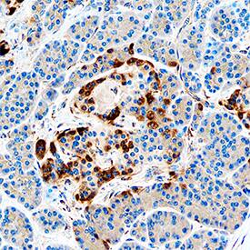
IGFBP-rp1/IGFBP-7 was detected in immersion fixed paraffin-embedded sections of human pancreas using Goat Anti-Human IGFBP-rp1/IGFBP-7 Antigen Affinity-purified Polyclonal Antibody (Catalog # AF1334) at 0.3 µg/mL overnight at 4 °C. Tissue was stained using the Anti-Goat HRP-DAB Cell & Tissue Staining Kit (brown; Catalog # CTS008) and counterstained with hematoxylin (blue). Specific staining was localized to islets. View our protocol for Chromogenic IHC Staining of Paraffin-embedded Tissue Sections.Applications for Human IGFBP-rp1/IGFBP-7 Antibody
Immunohistochemistry
Sample: Immersion fixed paraffin-embedded sections of human small intestine and human pancreas
Western Blot
Sample: HepG2 human hepatocellular carcinoma cell line
Formulation, Preparation, and Storage
Purification
Reconstitution
Formulation
Shipping
Stability & Storage
- 12 months from date of receipt, -20 to -70 °C as supplied.
- 1 month, 2 to 8 °C under sterile conditions after reconstitution.
- 6 months, -20 to -70 °C under sterile conditions after reconstitution.
Background: IGFBP-rp1/IGFBP-7
IGFBP-rp1, also known as Mac25/Angiomodulin (AGM), tumor-derived adhesion factor (TAF) and prostacyclin-stimulating factor (PSF), is a secreted protein that contains three protein domain modules. Human IGFBP-rp1 cDNA encodes 282 amino acid (aa) residue precursor protein with a putative 26 aa signal peptide. Mature IGFBP-rp1 is a glycosylated protein with an N-terminal IGFBP domain, followed by a Kazal-type serine proteinase inhibitor domain and a C-terminal immunoglobulin-like C2-type domain. The similarity of IGFBP-rp1 with the IGFBPs is confined to the N-terminal IGFBP domain, which contains all 12 of the conserved cysteine residues found in IGFBP-1 through 5. Human and mouse IGFBP-rp1 are highly homologous. Discounting a segment of 43 aa near the N-terminus that is missing in the mouse homologue, human and mouse IGFBP-rp1 share 94% aa sequence identity. IGFBP-rp1 is expressed in many normal tissues and in cancer cells. It is abundantly expressed in high endothelial venules (HEVs) of blood vessels in the secondary lymphoid tissues. The expression of IGFBP-rp1 is upregulated in senescing epithelial cells and by retinoic acid. IGFBP-rp1 binds IGF and insulin with very low affinity and has been shown to enhance the mitogenic actions of IGF and insulin. IGFBP-rp1 also has IGF/insulin-independent activities. It interacts with heparan sulfate proteoglycans, type IV collagen, and specific chemokines. IGFBP‑rp1 supports weak cell adhesion, promotes cell spreading on type IV collagen, and stimulates the production of the potent vasodilator PGI2. It modulates tumor cell growth and has also been implicated in angiogenesis. IGFBP-rp1 is proteolytically cleaved between lysine 97 and alanine 98. Cleaved IGFBP-rp1 has enhanced cell attachment activity but can no longer bind IGF/insulin (1-3).
References
- Hwa, V. et al. (1999) Endocrinology Rev. 20:761.
- Nagakubo, D. et al. (2003) J. Immunol. 171:553.
- Ahmed, S. et al. (2003) Biochem. Biophys. Res. Commun. 310:612.
Long Name
Alternate Names
Gene Symbol
UniProt
Additional IGFBP-rp1/IGFBP-7 Products
Product Documents for Human IGFBP-rp1/IGFBP-7 Antibody
Product Specific Notices for Human IGFBP-rp1/IGFBP-7 Antibody
For research use only
