Human IL-17/IL-17A Antibody
R&D Systems, part of Bio-Techne | Catalog # AF-317-NA


Key Product Details
Validated by
Species Reactivity
Validated:
Cited:
Applications
Validated:
Cited:
Label
Antibody Source
Product Specifications
Immunogen
Ile20-Ala155
Accession # Q16552
Specificity
Clonality
Host
Isotype
Endotoxin Level
Scientific Data Images for Human IL-17/IL-17A Antibody
Detection of Human IL‑17 by Western Blot.
Western blot shows lysates of human primary naïve CD4+ T cells, along with whole cell lysates (WCL) and conditioned-media supernatant (Supe) of human primary differentiated Th17 cells. PVDF membrane was probed with 1 µg/mL of Goat Anti-Human IL-17 Antigen Affinity-purified Polyclonal Antibody (Catalog # AF-317-NA) followed by HRP-conjugated Anti-Goat IgG Secondary Antibody (HAF017). A specific band was detected for IL-17 at approximately 15 kDa (as indicated). This experiment was conducted under reducing conditions and using Immunoblot Buffer Group 1.IL‑17/IL‑17A in Human Tonsil.
IL-17/IL-17A was detected in immersion fixed paraffin-embedded sections of human tonsil using Goat Anti-Human IL-17/IL-17A Antigen Affinity-purified Polyclonal Antibody (Catalog # AF-317-NA) at 1 µg/mL for 1 hour at room temperature followed by incubation with the Anti-Goat IgG VisUCyte™ HRP Polymer Antibody (VC004). Tissue was stained using DAB (brown) and counterstained with hematoxylin (blue). Specific staining was localized to lymphocytes. View our protocol for IHC Staining with VisUCyte HRP Polymer Detection Reagents.Immunoprecipitation of Human IL‑17.
Human IL-17 was immunoprecipitated from 100 µg of human primary differentiated Th17 cell lysate following incubation with 3 µg Goat Anti-Human IL-17 Antigen Affinity-purified Polyclonal Antibody (Catalog # AF-317-NA) or control antibody (AB-108-C) overnight at 4 °C. IL-17-antibody complexes were absorbed using Protein G Sepharose. Immunoprecipitated IL-17 was detected by Western blot using 1 µg/mL Goat Anti-Human IL-17 Antigen Affinity-purified Polyclonal Antibody (Catalog # AF-317-NA). View our recommended buffer recipes for immunoprecipitation.Applications for Human IL-17/IL-17A Antibody
CyTOF-ready
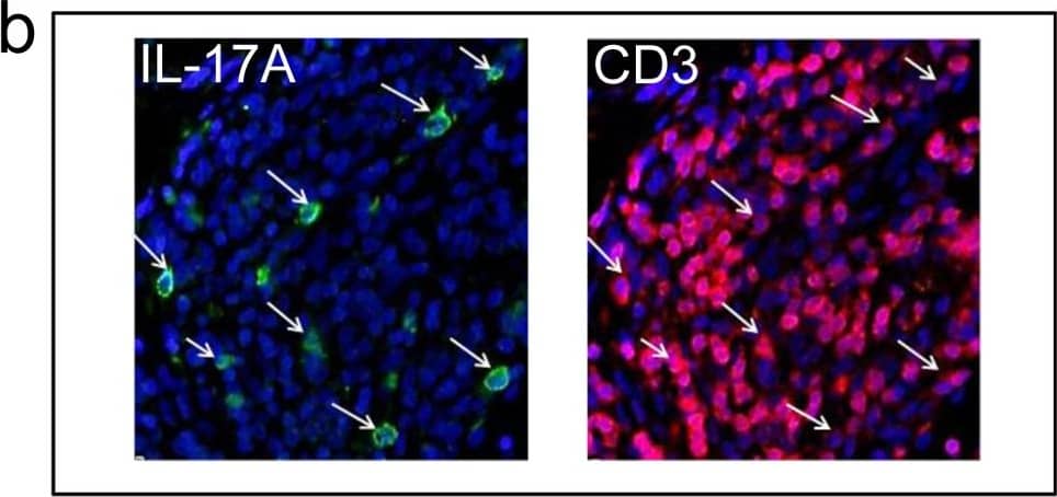
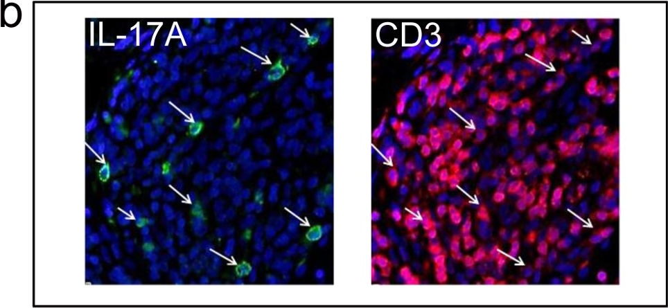
Immunocytochemistry
Sample: Immersion fixed human peripheral blood mononuclear cells treated with PHA
Immunohistochemistry
Sample: Immersion fixed paraffin-embedded sections of human tonsil
Immunoprecipitation
Sample: Human primary differentiated Th17 cells
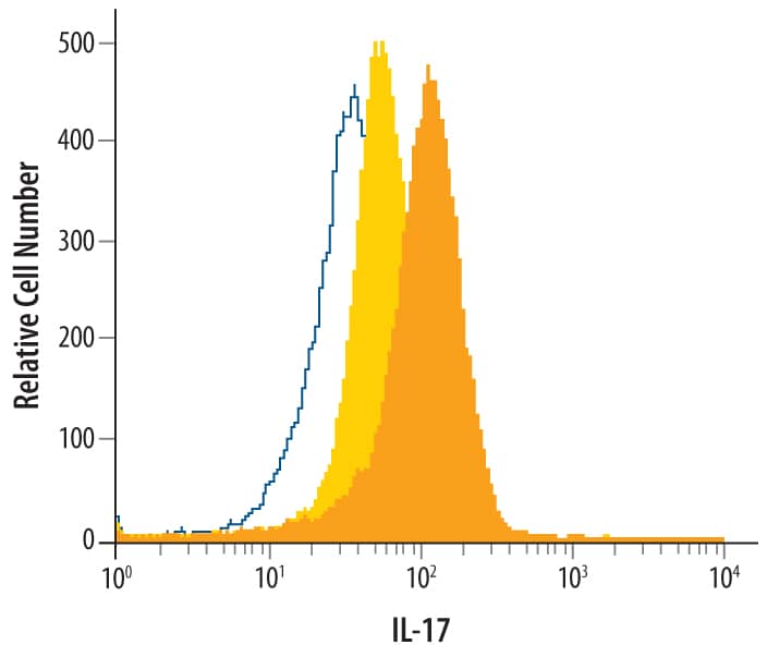
Intracellular Staining by Flow Cytometry
Sample: Human peripheral blood mononuclear cells treated with PMA and Ca2+ ionomycin, fixed with paraformaldehyde, and permeabilized with saponin
Western Blot
Sample: Human primary naïve CD4+ T cells and human primary differentiated Th17 cells
Neutralization
Reviewed Applications
Read 9 reviews rated 4.2 using AF-317-NA in the following applications:
Formulation, Preparation, and Storage
Purification
Reconstitution
Formulation
Shipping
Stability & Storage
- 12 months from date of receipt, -20 to -70 °C as supplied.
- 1 month, 2 to 8 °C under sterile conditions after reconstitution.
- 6 months, -20 to -70 °C under sterile conditions after reconstitution.
Background: IL-17/IL-17A
Interleukin 17 (also known as CTLA-8) is a T cell-expressed pleiotropic cytokine that exhibits a high degree of homology to a protein encoded by the ORF13 gene of herpesvirus Saimiri. cDNA clones encoding IL-17 have been isolated from activated rat, mouse and human T cells. Human IL-17 cDNA encodes a 155 amino acid (aa) residue precursor protein with a 19 amino acid residue signal peptide that is cleaved to yield the 136 aa residue mature IL-17 containing one potential N-linked glycosylation site. Both recombinant and natural IL-17 have been shown to exist as disulfide linked homodimers. At the amino acid level, human IL-17 shows 72% and 63% sequence identity with herpesvirus and rat IL-17, respectively. An IL-17 specific mouse cell surface receptor (IL-17 R) has recently been cloned. While the expression of IL-17 mRNA is restricted to activated T cells, the expression of mIL-17 R mRNA has been detected in virtually all cells and tissues tested. IL-17 exhibits multiple biological activities on a variety of cells including the induction of IL-6 and IL-8 production in fibroblasts, the enhancement of surface expression of ICAM-1 in fibroblasts, activation of NF-kappa B and costimulation of T cell proliferation.
Long Name
Alternate Names
Entrez Gene IDs
Gene Symbol
UniProt
Additional IL-17/IL-17A Products
Product Documents for Human IL-17/IL-17A Antibody
Product Specific Notices for Human IL-17/IL-17A Antibody
For research use only


