Human Indoleamine 2,3-dioxygenase/IDO Alexa Fluor® 488-conjugated Antibody
R&D Systems, part of Bio-Techne | Catalog # IC6030G


Key Product Details
Validated by
Species Reactivity
Validated:
Cited:
Applications
Validated:
Cited:
Label
Antibody Source
Product Specifications
Immunogen
Ala2-Gly403
Accession # P14902
Specificity
Clonality
Host
Isotype
Scientific Data Images
Detection of Indoleamine 2,3‑dioxygenase/IDO in Human Monocytes by Flow Cytometry.
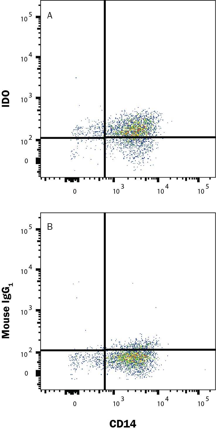
Human Monocytes were selected from PBMC using MagCellect Human CD14+ Cell Isolation Kit (MAGH105) and cultured overnight with Recombinant Human MCSF (50 ng/mL; 216-MC), Recombinant Human IFN gamma (50 ng/mL; 285-IF) and 50 ng/mL LPS and stained with (A) Mouse Anti-Human Indoleamine 2,3-dioxygenase/IDO Alexa Fluor® 488-conjugated Monoclonal Antibody (Catalog # IC6030G) or (B) Mouse IgG1 isotype control antibody (IC002G) and Mouse Anti-Human CD14 PE-conjugated Monoclonal Antibody (FAB3832P). To facilitate intracellular staining, cells were fixed and permeabilized with FlowX FoxP3/Transcription Factor Fixation & Perm Kit (FC012). Staining was performed using our Staining Intracellular Molecules protocol.Detection of Indoleamine 2,3‑dioxygenase/IDO in Human MDSCs by Flow Cytometry.
Human myeloid-derived suppressor cells (MDSCs) treated with 10 ng/mL Recombinant Human IL-6 (Catalog # 206-IL) and 10 ng/mL Recombinant Human GM-CSF (Catalog # 215-GM) for 7 days were stained with Mouse Anti-Human Siglec-3/CD33 APC-conjugated Monoclonal Antibody (Catalog # FAB1137A) and either (A) Mouse Anti-Human Indoleamine 2,3-dioxygenase/IDO Alexa Fluor® 488-conjugated Monoclonal Antibody (Catalog # IC6030G) or (B) Mouse IgG1Alexa Fluor 488 Isotype Control (Catalog # IC002G). To facilitate intracellular staining, cells were fixed with Flow Cytometry Fixation Buffer (Catalog # FC004) and permeabilized with Flow Cytometry Permeabilization/Wash Buffer I (Catalog # FC005). View our protocol for Staining Intracellular Molecules.Applications
Intracellular Staining by Flow Cytometry
Sample: Human myeloid-derived suppressor cells (MDSCs) treated with Recombinant Human IL‑6 (Catalog # 206-IL) and Recombinant Human GM‑CSF (Catalog # 215-GM) were fixed with Flow Cytometry Fixation Buffer (Catalog # FC004) and permeabilized with Flow Cytometry Permeabilization/Wash Buffer I (Catalog # FC005) and Human Monocytes selected from PBMC using MagCellect Human CD14+ Cell Isolation Kit (Catalog # MAGH105) and cultured overnight with Recombinant Human MCSF (50 ng/mL; Catalog # 216-MC), Recombinant Human IFN gamma (50 ng/mL; Catalog # 285-IF) and 50 ng/mL LPS
Reviewed Applications
Read 1 review rated 5 using IC6030G in the following applications:
Formulation, Preparation, and Storage
Purification
Formulation
Shipping
Stability & Storage
- 12 months from date of receipt, 2 to 8 °C as supplied.
Background: Indoleamine 2,3-dioxygenase/IDO
References
- Lewis-Ballester, A. et al. (2009) Proc. Natl. Acad. Sci. USA. 106:17371.
- Costantino, G. (2009) Expert Opin. Ther. Targets 13:247.
- Xu, H. et al. (2008) Immunol. Lett. 121:1.
- Lob, S. et al. (2009) Nat. Rev. Cancer 9:445.
- Curti, A. et al. (2009) Blood 113:2394.
Alternate Names
Gene Symbol
UniProt
Additional Indoleamine 2,3-dioxygenase/IDO Products
Product Specific Notices
This product is provided under an agreement between Life Technologies Corporation and R&D Systems, Inc, and the manufacture, use, sale or import of this product is subject to one or more US patents and corresponding non-US equivalents, owned by Life Technologies Corporation and its affiliates. The purchase of this product conveys to the buyer the non-transferable right to use the purchased amount of the product and components of the product only in research conducted by the buyer (whether the buyer is an academic or for-profit entity). The sale of this product is expressly conditioned on the buyer not using the product or its components (1) in manufacturing; (2) to provide a service, information, or data to an unaffiliated third party for payment; (3) for therapeutic, diagnostic or prophylactic purposes; (4) to resell, sell, or otherwise transfer this product or its components to any third party, or for any other commercial purpose. Life Technologies Corporation will not assert a claim against the buyer of the infringement of the above patents based on the manufacture, use or sale of a commercial product developed in research by the buyer in which this product or its components was employed, provided that neither this product nor any of its components was used in the manufacture of such product. For information on purchasing a license to this product for purposes other than research, contact Life Technologies Corporation, Cell Analysis Business Unit, Business Development, 29851 Willow Creek Road, Eugene, OR 97402, Tel: (541) 465-8300. Fax: (541) 335-0354.
For research use only
