Human Integrin alphaV beta5 Antibody
R&D Systems, part of Bio-Techne | Catalog # MAB2528


Conjugate
Catalog #
Key Product Details
Species Reactivity
Validated:
Human
Cited:
Human
Applications
Validated:
Adhesion Blockade, CyTOF-ready, Flow Cytometry, Immunocytochemistry, Immunoprecipitation
Cited:
Flow Cytometry, Immunocytochemistry, Immunohistochemistry, Neutralization
Label
Unconjugated
Antibody Source
Monoclonal Mouse IgG1 Clone # P5H9
Product Specifications
Immunogen
HT1080 human fibrosarcoma cell line
Specificity
Detects human Integrin alphaV beta5. Recognizes the human Integrin alphaV beta5 heterodimer and does not recognize the alphaV subunit in association with any other beta subunits.
Clonality
Monoclonal
Host
Mouse
Isotype
IgG1
Endotoxin Level
<0.10 EU per 1 μg of the antibody by the LAL method.
Scientific Data Images for Human Integrin alphaV beta5 Antibody
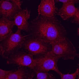
Integrin alphaV beta5 in HT1080 Human Cell Line.
Integrin aV beta5 was detected in immersion fixed HT1080 human fibrosarcoma cell line using Human Integrin aV beta5 Monoclonal Antibody (Catalog # MAB2528) at 10 µg/mL for 3 hours at room temperature. Cells were stained using the NorthernLights™ 557-conjugated Anti-Mouse IgG Secondary Antibody (red; Catalog # NL007) and counterstained with DAPI (blue). View our protocol for Fluorescent ICC Staining of Cells on Coverslips.Detection of Human Integrin alpha V beta 5 by Immunocytochemistry/Immunofluorescence
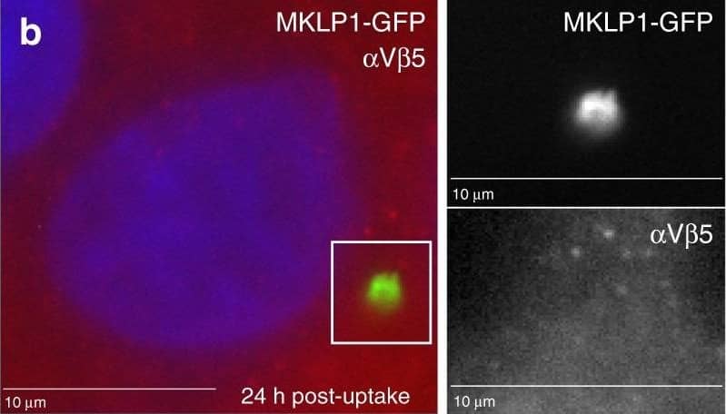
alphaV beta3 and EGFR mediate MBsome signaling. a–d HeLa cells were incubated with purified GFP-MBs for 3 h, followed by wash and another incubation for 24 h. Cells were then fixed and stained with anti-alpha V beta3 (a), anti-alpha V beta5 (b), anti-phospho-FAK (d) antibodies. Panels in c show control staining where primary antibodies were not added. Boxed regions mark the part of the image shown as a higher magnification image in the insets on the right. Scale bar is equivalent to 1 μm. e HeLa cells were co-incubated with purified GFP-MBs and EGF-Alexa647, followed by wash and another incubation for 24 h. Cells were then fixed and colocalization between MBs and EGF analyzed. Arrows point to MBsomes. f HeLa cells were co-incubated with purified GFP-MBs and non-labeled EGF, followed by wash and another incubation for 24 h. Cells were then fixed and stained with anti-phospho-EGFR antibodies. Arrows point to MBsomes. Scale bars in insets are equivalent to 2 μm. g HeLa cells were incubated with purified GFP-MBs. Cells were then washed and flow sorted to separate fractions with or without internalized GF-MBs. Equal number of cells from each fraction were then plated and incubated for 48 h in the presence or absence of 10 μm of EGFR inhibitor (erlotinib). Cells were then washed again and incubated for another 48 h followed by cell counting to determine the number of cells. Data shown are the means and standard deviations derived from three independent experiments (one-way ANOVA) Image collected and cropped by CiteAb from the following publication (https://pubmed.ncbi.nlm.nih.gov/31320617), licensed under a CC-BY license. Not internally tested by R&D Systems.Applications for Human Integrin alphaV beta5 Antibody
Application
Recommended Usage
Adhesion Blockade
Wayner, E.A. et al. (1991) J. Cell Biol. 113:919.
CyTOF-ready
Ready to be labeled using established conjugation methods. No BSA or other carrier proteins that could interfere with conjugation.
Flow Cytometry
2.5 µg/106 cells
Sample: MCF-7 human breast cancer cell line
Sample: MCF-7 human breast cancer cell line
Immunocytochemistry
8-25 µg/mL
Sample: Immersion fixed HT1080 human fibrosarcoma cell line, M21 human melanoma cell line, and H2981 human lung carcinoma cell line
Sample: Immersion fixed HT1080 human fibrosarcoma cell line, M21 human melanoma cell line, and H2981 human lung carcinoma cell line
Immunoprecipitation
Wayner, E.A. et al. (1991) J. Cell Biol. 113:919.
Reviewed Applications
Read 1 review rated 5 using MAB2528 in the following applications:
Formulation, Preparation, and Storage
Purification
Protein A or G purified from hybridoma culture supernatant
Reconstitution
Reconstitute at 0.5 mg/mL in sterile PBS. For liquid material, refer to CoA for concentration.
Formulation
Lyophilized from a 0.2 μm filtered solution in PBS with Trehalose. *Small pack size (SP) is supplied either lyophilized or as a 0.2 µm filtered solution in PBS.
Shipping
Lyophilized product is shipped at ambient temperature. Liquid small pack size (-SP) is shipped with polar packs. Upon receipt, store immediately at the temperature recommended below.
Stability & Storage
Use a manual defrost freezer and avoid repeated freeze-thaw cycles.
- 12 months from date of receipt, -20 to -70 °C as supplied.
- 1 month, 2 to 8 °C under sterile conditions after reconstitution.
- 6 months, -20 to -70 °C under sterile conditions after reconstitution.
Background: Integrin alpha V beta 5
Alternate Names
CD51, integrin subunit alpha V, ITGAV, MSK8, VNRA, VTNR
Entrez Gene IDs
3685 (Human)
Gene Symbol
ITGAV
Additional Integrin alpha V beta 5 Products
Product Documents for Human Integrin alphaV beta5 Antibody
Product Specific Notices for Human Integrin alphaV beta5 Antibody
For research use only
Loading...
Loading...
Loading...
Loading...
Loading...
