Human IRF3 Antibody
R&D Systems, part of Bio-Techne | Catalog # MAB4019


Key Product Details
Species Reactivity
Applications
Label
Antibody Source
Product Specifications
Immunogen
aa 206-427
Accession # Q14653
Specificity
Clonality
Host
Isotype
Scientific Data Images for Human IRF3 Antibody
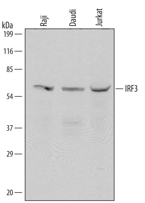
Detection of Human IRF3 by Western Blot.
Western blot shows lysates of Raji human Burkitt's lymphoma cell line, Daudi human Burkitt's lymphoma cell line, and Jurkat human acute T cell leukemia cell line. PVDF Membrane was probed with 1 µg/mL of Mouse Anti-Human IRF3 Monoclonal Antibody (Catalog # MAB4019) followed by HRP-conjugated Anti-Mouse IgG Secondary Antibody (Catalog # HAF007). A specific band was detected for IRF3 at approximately 60 kDa (as indicated). This experiment was conducted under reducing conditions and using Immunoblot Buffer Group 1.Detection of IRF3 in Daudi Human Cell Line by Flow Cytometry.
Daudi human Burkitt's lymphoma cell line was stained with Mouse Anti-Human IRF3 Monoclonal Antibody (Catalog # MAB4019, filled histogram) or isotype control antibody (Catalog # MAB0041, open histogram), followed by Phycoerythrin-conjugated Anti-Mouse IgG Secondary Antibody (Catalog # F0102B). To facilitate intracellular staining, cells were fixed with Flow Cytometry Fixation Buffer (Catalog # FC004) and permeabilized with Flow Cytometry Permeabilization/Wash Buffer I (Catalog # FC005).View our protocol for Staining Intracellular Molecules.Applications for Human IRF3 Antibody
CyTOF-ready
Intracellular Staining by Flow Cytometry
Sample: Daudi human Burkitt's lymphoma cell line fixed with Flow Cytometry Fixation Buffer (Catalog # FC004) and permeabilized with Flow Cytometry Permeabilization/Wash Buffer I (Catalog # FC005)
Western Blot
Sample: Raji human Burkitt's lymphoma cell line, Daudi human Burkitt's lymphoma cell line, and Jurkat human acute T cell leukemia cell line
Formulation, Preparation, and Storage
Purification
Reconstitution
Formulation
Shipping
Stability & Storage
- 12 months from date of receipt, -20 to -70 °C as supplied.
- 1 month, 2 to 8 °C under sterile conditions after reconstitution.
- 6 months, -20 to -70 °C under sterile conditions after reconstitution.
Background: IRF3
IRF3 (interferon response factor 3) is a 60 kDa member of the IRF family of proteins. Human IRF3 contains one DNA binding domain (aa 7‑107), a nuclear export signal (aa 139‑149) and multiple phosphorylation sites (aa 395‑407). Viral infection stimulates IRF3 phosphorylation, nuclear translocation and stimulation of IFN production. Alternate splice forms may exist. One will show a deletion of aa 201‑327, a second will show the same deletion plus an alternate start site at Met147, and a third will show a 125 aa substitution for the C-terminal 100 aa (aa 328‑427). Over aa 206‑427, human IRF3 is 76% and 83% aa identical to mouse and pig IRF3, respectively.
Long Name
Alternate Names
Gene Symbol
UniProt
Additional IRF3 Products
Product Documents for Human IRF3 Antibody
Product Specific Notices for Human IRF3 Antibody
For research use only
