Human LIF Antibody
R&D Systems, part of Bio-Techne | Catalog # AF-250-NA


Key Product Details
Species Reactivity
Validated:
Human
Cited:
Human, Primate - Macaca mulatta (Rhesus Macaque)
Applications
Validated:
Immunohistochemistry, Neutralization
Cited:
Immunocytochemistry, Immunohistochemistry, Immunohistochemistry-Paraffin, Neutralization, Western Blot
Label
Unconjugated
Antibody Source
Polyclonal Goat IgG
Product Specifications
Immunogen
E. coli-derived recombinant human LIF
Ser23-Phe202
Accession # P15018
Ser23-Phe202
Accession # P15018
Specificity
Detects human LIF in direct ELISAs.
Clonality
Polyclonal
Host
Goat
Isotype
IgG
Endotoxin Level
<0.10 EU per 1 μg of the antibody by the LAL method.
Scientific Data Images for Human LIF Antibody
LIF in Human Lung.
LIF was detected in immersion fixed paraffin-embedded sections of human lung array using Goat Anti-Human LIF Antigen Affinity-purified Polyclonal Antibody (Catalog # AF-250-NA) at 25 µg/mL overnight at 4 °C. Tissue was stained using the Anti-Goat HRP-DAB Cell & Tissue Staining Kit (brown; CTS008) and counterstained with hematoxylin (blue). Lower panel shows a lack of labeling if primary antibodies are omitted and tissue is stained only with secondary antibody followed by incubation with detection reagents. View our protocol for Chromogenic IHC Staining of Paraffin-embedded Tissue Sections.LIF in Human Alzheimer's Brain.
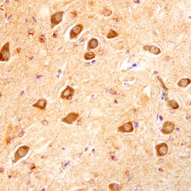
LIF was detected in immersion fixed paraffin-embedded sections of human Alzheimer's brain using Goat Anti-Human LIF Antigen Affinity-purified Polyclonal Antibody (Catalog # AF-250-NA) at 10 µg/mL overnight at 4 °C. Before incubation with the primary antibody, tissue was subjected to heat-induced epitope retrieval using Antigen Retrieval Reagent-Basic CTS013). Tissue was stained using the Anti-Goat HRP-DAB Cell & Tissue Staining Kit (brown CTS008) and counterstained with hematoxylin (blue). Specific staining was localized to neuronal cytoplasm. View our protocol for Chromogenic IHC Staining of Paraffin-embedded Tissue Sections.Cell Proliferation Induced by LIF and Neutralization by Human LIF Antibody.
Recombinant Human LIF (7734-LF) stimulates proliferation in the TF-1 human erythroleukemic cell line in a dose-depend-ent manner (orange line), as measured by Resazurin (AR002). Proliferation elicited by 3 ng/mL Recombinant Human LIF is neutralized (green line) by increasing concentrations of Goat Anti-Human LIF Antigen Affinity-purified Polyclonal Antibody (Catalog # AF-250-NA). The ND50 is typically 0.04-0.08 µg/mL.Applications for Human LIF Antibody
Application
Recommended Usage
Immunohistochemistry
5-15 µg/mL
Sample: Immersion fixed paraffin-embedded sections of human lung and human Alzheimer's brain subjected to heat-induced epitope retrieval using Antigen Retrieval Reagent-Basic (Catalog # CTS013).
Sample: Immersion fixed paraffin-embedded sections of human lung and human Alzheimer's brain subjected to heat-induced epitope retrieval using Antigen Retrieval Reagent-Basic (Catalog # CTS013).
Neutralization
Measured by its ability to neutralize LIF-induced proliferation in the TF‑1 human erythroleukemic cell line. Kitamura, T. et al. (1989) J. Cell Physiol. 140:323. The Neutralization Dose (ND50) is typically 0.04-0.08 µg/mL in the presence of 3 ng/mL Recombinant Human LIF.
Reviewed Applications
Read 1 review rated 4 using AF-250-NA in the following applications:
Formulation, Preparation, and Storage
Purification
Antigen Affinity-purified
Reconstitution
Reconstitute at 0.2 mg/mL in sterile PBS. For liquid material, refer to CoA for concentration.
Formulation
Lyophilized from a 0.2 μm filtered solution in PBS with Trehalose. *Small pack size (SP) is supplied either lyophilized or as a 0.2 µm filtered solution in PBS.
Shipping
Lyophilized product is shipped at ambient temperature. Liquid small pack size (-SP) is shipped with polar packs. Upon receipt, store immediately at the temperature recommended below.
Stability & Storage
Use a manual defrost freezer and avoid repeated freeze-thaw cycles.
- 12 months from date of receipt, -20 to -70 °C as supplied.
- 1 month, 2 to 8 °C under sterile conditions after reconstitution.
- 6 months, -20 to -70 °C under sterile conditions after reconstitution.
Background: LIF
Long Name
Leukemia Inhibitory Factor
Alternate Names
D Factor, Emfilermin, HILDA, MLPLI
Gene Symbol
LIF
UniProt
Additional LIF Products
Product Documents for Human LIF Antibody
Product Specific Notices for Human LIF Antibody
For research use only
Loading...
Loading...
Loading...
Loading...


