Human LYAR Antibody
R&D Systems, part of Bio-Techne | Catalog # AF6748

Key Product Details
Species Reactivity
Applications
Label
Antibody Source
Product Specifications
Immunogen
Lys288-Lys379
Accession # Q9NX58
Specificity
Clonality
Host
Isotype
Scientific Data Images for Human LYAR Antibody
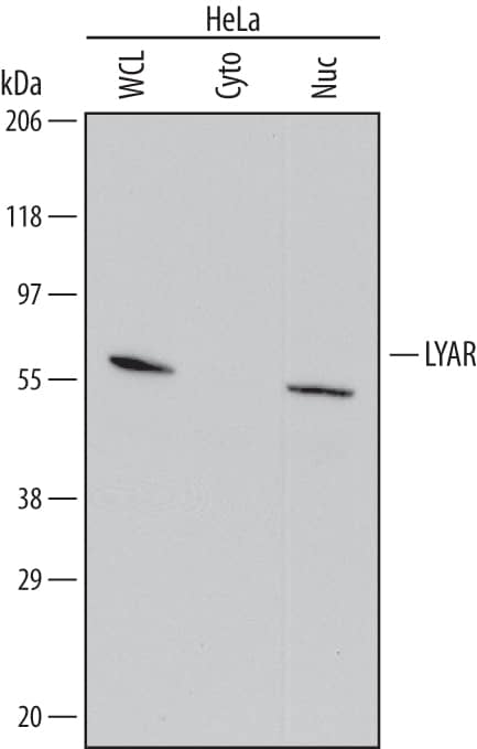
Detection of Human LYAR by Western Blot.
Western blot shows lysates of HeLa human cervical epithelial carcinoma cell line. Gels were loaded with 30 µg of whole cell lysate (WCL), 20 µg of cytoplasmic (Cyto), and 10 µg of nuclear extracts (Nuc). PVDF Membrane was probed with 0.1 µg/mL of Sheep Anti-Human LYAR Antigen Affinity-purified Polyclonal Antibody (Catalog # AF6748) followed by HRP-conjugated Anti-Sheep IgG Secondary Antibody (Catalog # HAF016). A specific band was detected for LYAR at approximately 50 kDa (as indicated). This experiment was conducted under reducing conditions and using Immunoblot Buffer Group 1.LYAR in BG01V Human Stem Cells.
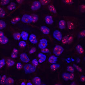
LYAR was detected in immersion fixed BG01V human embryonic stem cells using Sheep Anti-Human LYAR Antigen Affinity-purified Polyclonal Antibody (Catalog # AF6748) at 5 µg/mL for 3 hours at room temperature. Cells were stained using the NorthernLights™ 557-conjugated Anti-Sheep IgG Secondary Antibody (red; Catalog # NL010) and counterstained with DAPI (blue). Specific staining was localized to nucleoli. View our protocol for Fluorescent ICC Staining of Cells on Coverslips.Applications for Human LYAR Antibody
Immunocytochemistry
Sample: Immersion fixed BG01V human embryonic stem cells
Western Blot
Sample: HeLa human cervical epithelial carcinoma cell line
Formulation, Preparation, and Storage
Purification
Reconstitution
Formulation
Shipping
Stability & Storage
- 12 months from date of receipt, -20 to -70 °C as supplied.
- 1 month, 2 to 8 °C under sterile conditions after reconstitution.
- 6 months, -20 to -70 °C under sterile conditions after reconstitution.
Background: LYAR
LYAR (Ly-1/CD5 antibody reactive clone) is a 45-50 kDa nucleolar protein that was named for the ability of its antibody to cross-react with Ly-1. Its function is unclear; it is known to associate with MYCN and RRP1B, the latter association giving rise to the suggestion that LYAR is involved with RNA metabolism. Human LYAR is 379 amino acids (aa) in length. It contains two C2H2-type Zn finger regions (aa 6-25 and 33-51) followed by one coiled-coil region (aa 175-219) and an NLS (aa 217-222). There are multiple Zn-binding sites and three utilized phosphorylation sites at Ser244, Ser258 and Ser276. Over aa 288-379, human LYAR shares 75% aa identity with mouse LYAR.
Long Name
Alternate Names
Gene Symbol
UniProt
Additional LYAR Products
Product Documents for Human LYAR Antibody
Product Specific Notices for Human LYAR Antibody
For research use only

