Human Lymphotoxin-alpha /TNF-beta Antibody
R&D Systems, part of Bio-Techne | Catalog # MAB621R

Key Product Details
Species Reactivity
Applications
Label
Antibody Source
Product Specifications
Immunogen
Specificity
Clonality
Host
Isotype
Endotoxin Level
Scientific Data Images for Human Lymphotoxin-alpha /TNF-beta Antibody
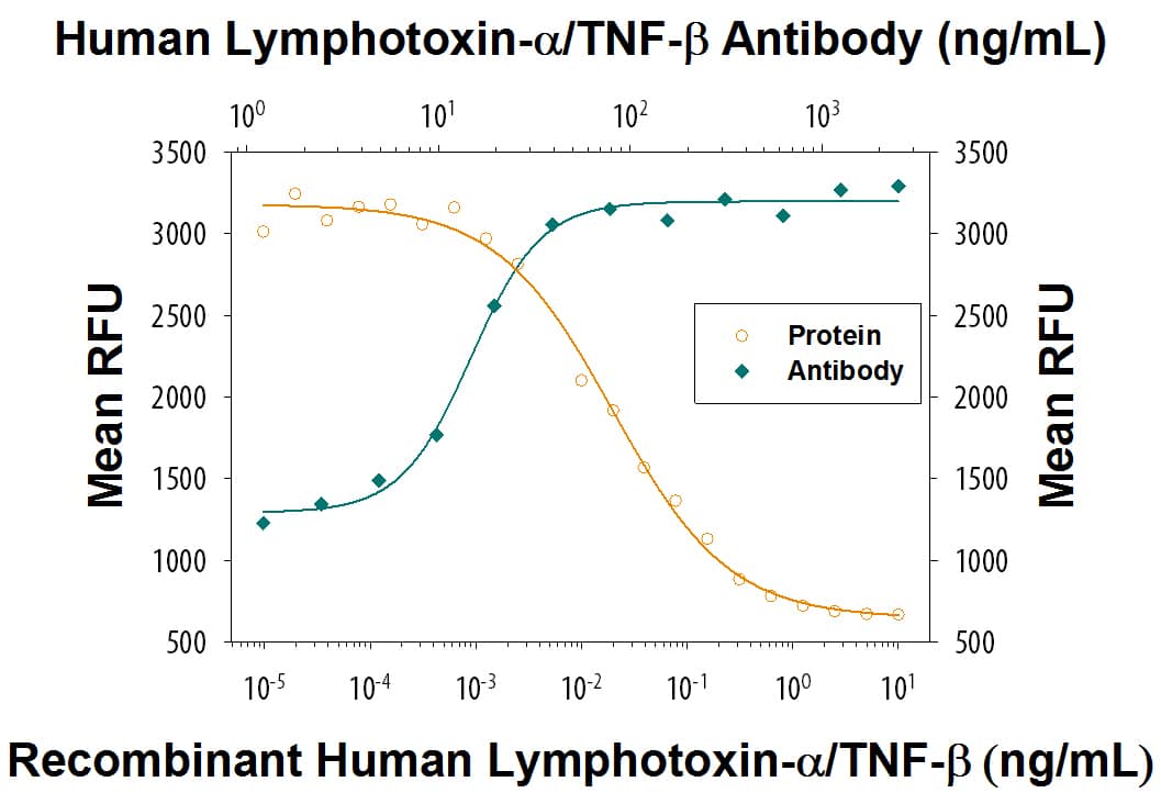
Cytotoxicity Induced by Lymphotoxin-alpha /TNF-beta and Neutral-ization by Human Lymphotoxin-alpha /TNF-beta Antibody.
Recombinant Human Lymphotoxin-a/TNF-beta (Catalog # 211-TBB) induces cytotoxicity in the the L-929 mouse fibroblast cell line in a dose-dependent manner (orange line), as measured by Resazurin (Catalog # AR002). Cytotoxicity elicited by Recombinant Human Lymphotoxin-a/TNF-beta (0.25 ng/mL) is neutralized (green line) by increasing concentrations of Mouse Anti-Human Lymphotoxin-a/TNF-beta Monoclonal Antibody (Catalog # MAB621R). The ND50 is typically 8-48 ng/mL in the presence of the metabolic inhibitor actinomycin D.Applications for Human Lymphotoxin-alpha /TNF-beta Antibody
Neutralization
Human Lymphotoxin-alpha /TNF-beta Sandwich Immunoassay
Formulation, Preparation, and Storage
Purification
Reconstitution
Formulation
Shipping
Stability & Storage
- 12 months from date of receipt, -20 to -70 °C as supplied.
- 1 month, 2 to 8 °C under sterile conditions after reconstitution.
- 6 months, -20 to -70 °C under sterile conditions after reconstitution.
Background: Lymphotoxin-alpha/TNF-beta
Tumor necrosis factor beta (TNF-beta), also known as lymphotoxin-alpha (LT-alpha), and TNF-alpha, are two structurally and functionally related proteins that bind to the same cell surface receptors (TNF RI and TNF RII) and produce a vast range of similar, but not identical, effects. Among these effects is the ability to kill certain tumor cells directly, from which the names tumor necrosis factor and lymphotoxin both derive. Mature TNF-beta /LT-alpha and TNF-alpha share approximately 35% protein sequence homology and the biologically active secreted forms of both proteins are homotrimers. Whereas TNF-alpha can exist as a type II membrane protein, TNF-beta /LT-alpha possesses a typical signal peptide sequence and is a secreted protein. It has been shown that TNF-beta /LT-alpha is also present on the cell surface of activated T, B and LAK cells as a heteromeric complex with LT-beta, a type II membrane protein that is another member of the TNF ligand family. The genes for TNF-alpha, TNF-beta /LT-alpha, and LT-beta are closely linked within the major histocompatibility complex.
TNF-beta /LT-alpha is expressed in activated T- and B-lymphocytes. In addition to its cytotoxic action on tumor cells, TNF-beta /LT-alpha has been shown to be a mediator of inflammation and immune function. Evidence is also accumulating that TNF-beta /LT-alpha and TNF-alpha are mediators in the pathogenesis of certain autoimmune diseases. TNF-beta /LT-alpha has also been shown to have a role in lymphoid organ development. Human and mouse TNF-beta /LT-alpha share approximately 74% homology in their amino acid sequence and exhibit cross-species activity.
Long Name
Alternate Names
Gene Symbol
Additional Lymphotoxin-alpha/TNF-beta Products
Product Documents for Human Lymphotoxin-alpha /TNF-beta Antibody
Product Specific Notices for Human Lymphotoxin-alpha /TNF-beta Antibody
For research use only
