Human MANF Antibody
R&D Systems, part of Bio-Techne | Catalog # AF3748

Key Product Details
Validated by
Knockout/Knockdown
Species Reactivity
Validated:
Human
Cited:
Human, Mouse, Rat
Applications
Validated:
Knockout Validated, Simple Western, Western Blot
Cited:
ELISA Capture, Immunocytochemistry, Immunoprecipitation, Western Blot
Label
Unconjugated
Antibody Source
Polyclonal Goat IgG
Product Specifications
Immunogen
Mouse myeloma cell line NS0-derived recombinant human MANF
Leu22-Leu179
Accession # P55145
Leu22-Leu179
Accession # P55145
Specificity
Detects human MANF in direct ELISAs and Western blots.
Clonality
Polyclonal
Host
Goat
Isotype
IgG
Scientific Data Images for Human MANF Antibody
Detection of Human MANF by Western Blot.
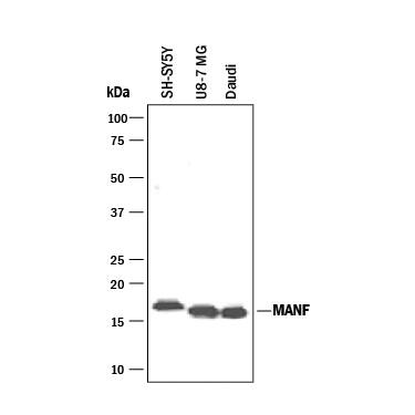
Western blot shows lysates of SH-SY5Y human neuroblastoma cell line, U-87 MG human glioblastoma/astrocytoma cell line, and Daudi human Burkitt's lymphoma cell line. PVDF membrane was probed with 1 µg/mL of Goat Anti-Human MANF Antigen Affinity-purified Polyclonal Antibody (Catalog # AF3748) followed by HRP-conjugated Anti-Goat IgG Secondary Antibody (HAF017). A specific band was detected for MANF at approximately 17 kDa (as indicated). This experiment was conducted under reducing conditions and using Western Blot Buffer Group 1.Detection of Human MANF by Simple WesternTM.
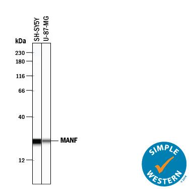
Simple Western lane view shows lysates of SH-SY5Y human neuroblastoma cell line and U-87 MG human glioblastoma/astrocytoma cell line, loaded at 0.2 mg/mL. A specific band was detected for MANF at approximately 24 kDa (as indicated) using 10 µg/mL of Goat Anti-Human MANF Antigen Affinity-purified Polyclonal Antibody (Catalog # AF3748) followed by 1:50 dilution of HRP-conjugated Anti-Goat IgG Secondary Antibody (Catalog # HAF109). This experiment was conducted under reducing conditions and using the 12-230 kDa separation system.Western Blot Shows Human MANF Specificity by Using Knockout Cell Line.
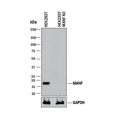
Western blot shows lysates of HEK293T human embryonic kidney parental cell line and MANF knockout HEK293T cell line (KO). PVDF membrane was probed with 1 µg/mL of Goat Anti-Human MANF Antigen Affinity-purified Polyclonal Antibody (Catalog # AF3748) followed by HRP-conjugated Anti-Goat IgG Secondary Antibody (Catalog # HAF017). A specific band was detected for MANF at approximately 17 kDa (as indicated) in the parental HEK293T cell line, but is not detectable in knockout HEK293Tcell line. GAPDH (Catalog # AF5718) is shown as a loading control. This experiment was conducted under reducing conditions and using Immunoblot Buffer Group 1.Applications for Human MANF Antibody
Application
Recommended Usage
Knockout Validated
MANF
is specifically detected in HEK293T human embryonic kidney parental cell line but is not detectable in
MANF knockout HEK293T cell line.
Simple Western
10 µg/mL
Sample: SH‑SY5Y human neuroblastoma cell line and U‑87 MG human glioblastoma/astrocytoma cell line
Sample: SH‑SY5Y human neuroblastoma cell line and U‑87 MG human glioblastoma/astrocytoma cell line
Western Blot
1 µg/mL
Sample: SH‑SY5Y human neuroblastoma cell line, U‑87 MG human glioblastoma/astrocytoma cell line, and Daudi human Burkitt's lymphoma cell line
Sample: SH‑SY5Y human neuroblastoma cell line, U‑87 MG human glioblastoma/astrocytoma cell line, and Daudi human Burkitt's lymphoma cell line
Reviewed Applications
Read 3 reviews rated 4 using AF3748 in the following applications:
Formulation, Preparation, and Storage
Purification
Antigen Affinity-purified
Reconstitution
Reconstitute at 0.2 mg/mL in sterile PBS. For liquid material, refer to CoA for concentration.
Formulation
Lyophilized from a 0.2 μm filtered solution in PBS with Trehalose. *Small pack size (SP) is supplied either lyophilized or as a 0.2 µm filtered solution in PBS.
Shipping
Lyophilized product is shipped at ambient temperature. Liquid small pack size (-SP) is shipped with polar packs. Upon receipt, store immediately at the temperature recommended below.
Stability & Storage
Use a manual defrost freezer and avoid repeated freeze-thaw cycles.
- 12 months from date of receipt, -20 to -70 °C as supplied.
- 1 month, 2 to 8 °C under sterile conditions after reconstitution.
- 6 months, -20 to -70 °C under sterile conditions after reconstitution.
Background: MANF
References
- Petrova, P.S. et al. (2003) J. Mol. Neurosci. 20:173.
- Shridhar, V. et al. (1996) Oncogene 12:1931.
- Shridhar, R. et al. (1996) Cancer Res. 56:5576.
- Shridhar, V. et al. (1997) Oncogene 14:2213.
- Apostolou, A. et al. (2008) Exp. Cell Res. 314:2454.
- Tadimalla, A. et al. (2008) Circ. Res. 103:1249.
Long Name
Mesencephalic Astrocyte-derived Neurotrphic Factor
Alternate Names
ARMET, ARP
Gene Symbol
MANF
UniProt
Additional MANF Products
Product Documents for Human MANF Antibody
Product Specific Notices for Human MANF Antibody
For research use only
Loading...
Loading...
Loading...
Loading...


