Human Metadherin Antibody
R&D Systems, part of Bio-Techne | Catalog # AF7644

Key Product Details
Species Reactivity
Applications
Label
Antibody Source
Product Specifications
Immunogen
Lys168-Ser298
Accession # Q86UE4
Specificity
Clonality
Host
Isotype
Scientific Data Images for Human Metadherin Antibody
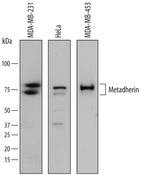
Detection of Human Metadherin by Western Blot.
Western blot shows lysates of MDA-MB-231 human breast cancer cell line, HeLa human cervical epithelial carcinoma cell line, and MDA-MB-453 human breast cancer cell line. PVDF membrane was probed with 0.2 µg/mL of Sheep Anti-Human Metadherin Antigen Affinity-purified Polyclonal Antibody (Catalog # AF7644) followed by HRP-conjugated Anti-Sheep IgG Secondary Antibody (Catalog # HAF016). Specific bands were detected for Metadherin at approximately 80 and 75 kDa (as indicated). This experiment was conducted under reducing conditions and using Immunoblot Buffer Group 1.Metadherin in HeLa Human Cell Line.
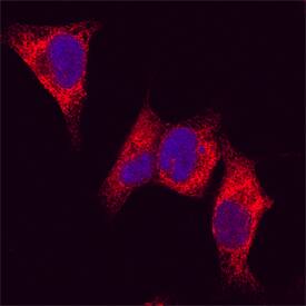
Metadherin was detected in immersion fixed HeLa human cervical epithelial carcinoma cell line using Sheep Anti-Human Metadherin Antigen Affinity-purified Polyclonal Antibody (Catalog # AF7644) at 5 µg/mL for 3 hours at room temperature. Cells were stained using the NorthernLights™ 557-conjugated Anti-Sheep IgG Secondary Antibody (red; Catalog # NL010) and counterstained with DAPI (blue). Specific staining was localized to cytoplasm. View our protocol for Fluorescent ICC Staining of Cells on Coverslips.Metadherin in Human Breast.
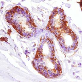
Metadherin was detected in immersion fixed paraffin-embedded sections of human breast using Sheep Anti-Human Metadherin Antigen Affinity-purified Polyclonal Antibody (Catalog # AF7644) at 3 µg/mL overnight at 4 °C. Tissue was stained using the Anti-Sheep HRP-DAB Cell & Tissue Staining Kit (brown; Catalog # CTS019) and counterstained with hematoxylin (blue). Specific staining was localized to cytoplasm and nuclei. View our protocol for Chromogenic IHC Staining of Paraffin-embedded Tissue Sections.Applications for Human Metadherin Antibody
Immunocytochemistry
Sample: Immersion fixed HeLa human cervical epithelial carcinoma cell line
Immunohistochemistry
Sample: Immersion fixed paraffin-embedded sections of human breast
Western Blot
Sample: MDA‑MB‑231 human breast cancer cell line, HeLa human cervical epithelial carcinoma cell line, and MDA‑MB‑453 human breast cancer cell line
Formulation, Preparation, and Storage
Purification
Reconstitution
Formulation
Shipping
Stability & Storage
- 12 months from date of receipt, -20 to -70 °C as supplied.
- 1 month, 2 to 8 °C under sterile conditions after reconstitution.
- 6 months, -20 to -70 °C under sterile conditions after reconstitution.
Background: Metadherin
MTDH (Metadherin; also LYRIC and Astrocyte Elevated Gene product-1/AEG1) is an 80 kDa membrane-bound protein that belongs to no known structural protein family. It has restricted expression, being found in breast ductal epithelium, striated muscle, select neurons and astrocytes, as well as multiple tumor types of diverse origin. MTDH is considered a mediator of Myc and Ha-Ras, and its activity is associated with stimulation of the beta-catenin/Wnt and NFkB signaling pathways. It is also believed to contribute to tight junction formation via an interaction with ZO-1. MTDH is also found in the nucleus where it complexes with CBP and p65. Human MTDH is 582 amino acids (aa) in length. It appears to be a type III transmembrane protein (N-terminus luminal/extracellular with no signal sequence) that is found embedded in both the plasma membrane and ER. It contains an N-terminal luminal region (aa 1-48), plus a large 513 aa cytoplasmic domain (aa 70-582) that possesses at least 11 utilized Ser/Thr phosphorylation sites. There is the potential for ubiquitination, SUMOylation, acetylation and proteolytic processing, and this may account for multiple bands in SDS-PAGE that represent MWs of 20, 35, 50-55, 70-75, and 86 kDa. Over aa 169-298, human MTDH shares 97% aa identity with mouse MDTH.
Alternate Names
Gene Symbol
UniProt
Additional Metadherin Products
Product Documents for Human Metadherin Antibody
Product Specific Notices for Human Metadherin Antibody
For research use only


