Human mGluR5 Antibody
R&D Systems, part of Bio-Techne | Catalog # MAB45141

Key Product Details
Species Reactivity
Validated:
Cited:
Applications
Validated:
Cited:
Label
Antibody Source
Product Specifications
Immunogen
Ser19-Ser509
Accession # P41594
Specificity
Clonality
Host
Isotype
Scientific Data Images for Human mGluR5 Antibody
mGluR5 in Human Brain.
mGluR5 was detected in immersion fixed paraffin-embedded sections of human brain (cerebellum) using 25 µg/mL Mouse Anti-Human mGluR5 Monoclonal Antibody (Catalog # MAB45141) overnight at 4 °C. Tissue was stained with the Anti-Mouse HRP-DAB Cell & Tissue Staining Kit (brown; Catalog # CTS002) and counterstained with hematoxylin (blue). View our protocol for Chromogenic IHC Staining of Paraffin-embedded Tissue Sections.Detection of Mouse mGluR5 by Immunocytochemistry/Immunofluorescence
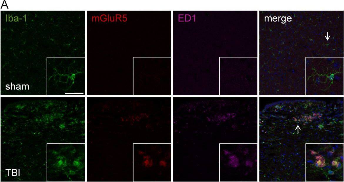
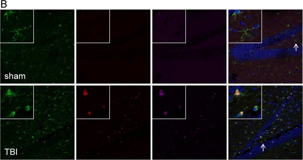
mGluR5 is expressed in chronically activated microglia at one month post TBI. (A and B) mGluR5 expression was evaluated in the cortex (A) and hippocampus (B) of sham or TBI brains at one month post-injury. Iba-1 (green) and ED1 (magenta) labeled activated microglia. mGluR5 expression (red) was undetectable in resting microglia that displayed a ramified cellular morphology, but was strongly up-regulated in highly reactive microglia that displayed a hypertrophic or bushy cellular morphology and co-expressed ED1 (merged). (C) The membrane bound component of the NADPH oxidase enzyme, gp91phox (red), co-localized (merged) with ED1-positive reactive microglial (green) at one month post-TBI. Bar = 25 μm. Image collected and cropped by CiteAb from the following publication (https://pubmed.ncbi.nlm.nih.gov/22373400), licensed under a CC-BY license. Not internally tested by R&D Systems.Detection of Human mGluR5 by Immunocytochemistry/Immunofluorescence
mGluR5 is expressed in chronically activated microglia at one month post TBI. (A and B) mGluR5 expression was evaluated in the cortex (A) and hippocampus (B) of sham or TBI brains at one month post-injury. Iba-1 (green) and ED1 (magenta) labeled activated microglia. mGluR5 expression (red) was undetectable in resting microglia that displayed a ramified cellular morphology, but was strongly up-regulated in highly reactive microglia that displayed a hypertrophic or bushy cellular morphology and co-expressed ED1 (merged). (C) The membrane bound component of the NADPH oxidase enzyme, gp91phox (red), co-localized (merged) with ED1-positive reactive microglial (green) at one month post-TBI. Bar = 25 μm. Image collected and cropped by CiteAb from the following publication (https://pubmed.ncbi.nlm.nih.gov/22373400), licensed under a CC-BY license. Not internally tested by R&D Systems.Applications for Human mGluR5 Antibody
Immunohistochemistry
Sample: Immersion fixed paraffin-embedded sections of human brain (cerebellum)
Formulation, Preparation, and Storage
Purification
Reconstitution
Formulation
Shipping
Stability & Storage
- 12 months from date of receipt, -20 to -70 °C as supplied.
- 1 month, 2 to 8 °C under sterile conditions after reconstitution.
- 6 months, -20 to -70 °C under sterile conditions after reconstitution.
Background: mGluR5
Human metabotropic glutamate receptor 5 (mGluR5; also known as mGluR5b) is a 150 kDa, 7-transmembrane glycoprotein that belongs to group I of the C-family of G-protein coupled receptors. mGluR5 is constitutively expressed and regulates neuronal ion channel activity. Human mGluR5 is 1212 amino acids (aa) in length and contains an N-terminal extracellular domain (ECD) of 558 aa. Through its ECD, mGluR5 either homodimerizes or heterodimerizes with the Ca++-sensor receptor. There is one alternate splice form (mGluR5a) that shows a 32 aa deletion between aa 877-908 in the cytoplasmic tail. Over aa 21-509, human mGluR5 shares 98% aa sequence identity with mouse, rat, and dog mGluR5.
Long Name
Alternate Names
Gene Symbol
UniProt
Additional mGluR5 Products
Product Documents for Human mGluR5 Antibody
Product Specific Notices for Human mGluR5 Antibody
For research use only


