Human MMP-2 Antibody
R&D Systems, part of Bio-Techne | Catalog # AF902


Key Product Details
Species Reactivity
Validated:
Cited:
Applications
Validated:
Cited:
Label
Antibody Source
Product Specifications
Immunogen
Ile34-Cys660
Accession # P08253
Specificity
Clonality
Host
Isotype
Scientific Data Images for Human MMP-2 Antibody
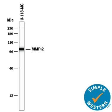
Detection of Human MMP-2 by Western Blot.
Western blot shows lysate of U-118-MG human glioblastoma/astrocytoma cell line. PVDF membrane was probed with 1 µg/mL of Goat Anti-Human MMP-2 Antigen Affinity-purified Polyclonal Antibody (Catalog # AF902) followed by HRP-conjugated Anti-Goat IgG Secondary Antibody (HAF017). A specific band was detected for MMP-2 at approximately 72 kDa (as indicated). This experiment was conducted under reducing conditions and using Immunoblot Buffer Group 1.MMP‑2 in Human Ovarian Cancer Tissue.
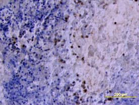
MMP-2 was detected in immersion fixed paraffin-embedded sections of human ovarian cancer tissue using Goat Anti-Human MMP-2 Antigen Affinity-purified Poly-clonal Antibody (Catalog # AF902) at 10 µg/mL overnight at 4 °C. Tissue was stained using the Anti-Goat HRP-DAB Cell & Tissue Staining Kit (brown; CTS008) and counter-stained with hematoxylin (blue). View our protocol for Chromogenic IHC Staining of Paraffin-embedded Tissue Sections.MMP‑2 in Human Ovary.
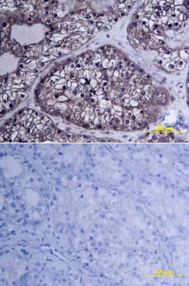
MMP-2 was detected in immersion fixed paraffin-embedded sections of human ovarian array using Goat Anti-Human MMP-2 Antigen Affinity-purified Polyclonal Antibody (Catalog # AF902) at 10 µg/mL overnight at 4 °C. Tissue was stained using the Anti-Goat HRP-DAB Cell & Tissue Staining Kit (brown; CTS008) and counterstained with hematoxylin (blue). Lower panel shows a lack of labeling if primary antibodies are omitted and tissue is stained only with secondary antibody followed by incubation with detection reagents. View our protocol for Chromogenic IHC Staining of Paraffin-embedded Tissue Sections.Applications for Human MMP-2 Antibody
Immunohistochemistry
Sample: Immersion fixed paraffin-embedded sections of human ovarian cancer tissue and normal human ovarian array
Immunoprecipitation
Sample: Conditioned cell culture medium spiked with Recombinant Human MMP-2 (Catalog # 902-MP), see our available Western blot detection antibodies
Simple Western
Sample: U‑118‑MG human glioblastoma/astrocytoma cell line
Western Blot
Sample: U‑118‑MG human glioblastoma/astrocytoma cell line
Reviewed Applications
Read 4 reviews rated 4.3 using AF902 in the following applications:
Formulation, Preparation, and Storage
Purification
Reconstitution
Formulation
Shipping
Stability & Storage
- 12 months from date of receipt, -20 to -70 °C as supplied.
- 1 month, 2 to 8 °C under sterile conditions after reconstitution.
- 6 months, -20 to -70 °C under sterile conditions after reconstitution.
Background: MMP-2
Matrix metalloproteinases are a family of zinc and calcium dependent endopeptidases with the combined ability to degrade all the components of the extracellular matrix. MMP-2 (gelatinase A), a type IV collagenase, can degrade a broad range of substrates including type IV, V, VII and X collagens as well as elastin and fibronectin. It is believed to act synergistically with interstitial collagenase (MMP-1) in the degradation of fibrillar collagens as it degrades their denatured gelatin forms. MMP-2 has been shown to be associated with many connective tissue cells as well as neutrophils, macrophages and monocytes. Structurally, MMP-2 may be divided into several distinct domains: a pro-domain which is cleaved upon activation; a catalytic domain containing the zinc binding site; a fibronectin-like domain thought to play a role in substrate targeting; and a carboxyl terminal (hemopexin-like) domain containing 2 N-linked glycosylation sites.
Long Name
Alternate Names
Gene Symbol
UniProt
Additional MMP-2 Products
Product Documents for Human MMP-2 Antibody
Product Specific Notices for Human MMP-2 Antibody
For research use only


