Human/Mouse Angiopoietin-1 Antibody
R&D Systems, part of Bio-Techne | Catalog # AF923


Key Product Details
Species Reactivity
Validated:
Cited:
Applications
Validated:
Cited:
Label
Antibody Source
Product Specifications
Immunogen
Ser20-Phe498
Accession # Q5HYA0
Specificity
Clonality
Host
Isotype
Scientific Data Images for Human/Mouse Angiopoietin-1 Antibody
Angiopoietin‑1 in Human Prostate Cancer Tissue.
Angiopoietin-1 was detected in immersion fixed paraffin-embedded sections of human prostate cancer tissue using 15 µg/mL Goat Anti-Human/Mouse Angiopoietin-1 Antigen Affinity-purified Polyclonal Antibody (Catalog # AF923) overnight at 4 °C. Tissue was stained with the Anti-Goat HRP-DAB Cell & Tissue Staining Kit (brown; CTS008) and counterstained with hematoxylin (blue). View our protocol for Chromogenic IHC Staining of Paraffin-embedded Tissue Sections.Angiopoietin‑1 in Human Prostate.
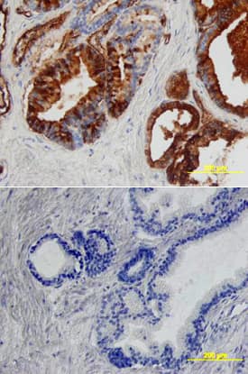
Angiopoietin-1 was detected in immersion fixed paraffin-embedded sections of human prostate array using Goat Anti-Human/Mouse Angiopoietin-1 Antigen Affinity-purified Polyclonal Antibody (Catalog # AF923) at 5 µg/mL overnight at 4 °C. Tissue was stained using the Anti-Goat HRP-DAB Cell & Tissue Staining Kit (brown; CTS008) and counterstained with hematoxylin (blue). Lower panel shows a lack of labeling if primary antibodies are omitted and tissue is stained only with secondary antibody followed by incubation with detection reagents. View our protocol for Chromogenic IHC Staining of Paraffin-embedded Tissue Sections.Angiopoietin‑1 in Mouse Embryo.
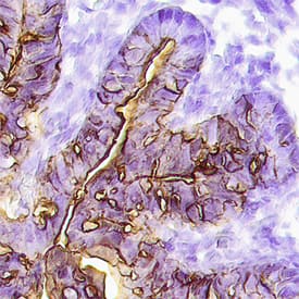
Angiopoietin-1 was detected in perfusion fixed frozen sections of mouse embryo (15 d.p.c.) using Goat Anti-Human/Mouse Angiopoietin-1 Antigen Affinity-purified Polyclonal Antibody (Catalog # AF923) at 15 µg/mL overnight at 4 °C. Tissue was stained using the Anti-Goat HRP-DAB Cell & Tissue Staining Kit (brown; CTS008) and counterstained with hematoxylin (blue). Specific staining was localized to endothelium in the loop of the midgut. View our protocol for Chromogenic IHC Staining of Frozen Tissue Sections.Applications for Human/Mouse Angiopoietin-1 Antibody
Immunohistochemistry
Sample: Immersion fixed paraffin-embedded sections of human prostate cancer tissue and normal human prostate tissue, and perfusion fixed frozen sections of mouse embryo (15 d.p.c.)
Western Blot
Sample: Recombinant Human Angiopoietin-1 (Catalog # 923-AN)
Reviewed Applications
Read 2 reviews rated 2.5 using AF923 in the following applications:
Formulation, Preparation, and Storage
Purification
Reconstitution
Formulation
Shipping
Stability & Storage
- 12 months from date of receipt, -20 to -70 °C as supplied.
- 1 month, 2 to 8 °C under sterile conditions after reconstitution.
- 6 months, -20 to -70 °C under sterile conditions after reconstitution.
Background: Angiopoietin-1
Angiopoietin-1 (Ang-1) and Angiopoietin-2 (Ang-2) are two closely related secreted ligands which bind with similar affinity to Tie-2, a receptor tyrosine kinase with immunoglobulin and epidermal growth factor homology domains expressed primarily on endothelial cells and early hematopoietic cells. Tie-2 and angiopoietins have been shown to play critical roles in embryogenic angiogenesis and in maintaining the integrity of the adult vasculature (1).
Ang-1 cDNA encodes a 498 amino acid (aa) residue precursor protein that contains a coiled-coiled domain near the amino-terminus and a fibrinogen-like domain at the C-terminus. Human Ang-1 shares approximately 97% and 60% amino acid sequence identity with mouse Ang-1 and human Ang-2, respectively (1, 2). Ang-1 activates Tie-2 signaling on endothelial cells to promote chemotaxis, cell survival, cell sprouting, vessel growth and stabilization (1, 3, 4). Ang-2 has alternatively been reported to be an antagonist for Ang-1 induced Tie-2 signaling as well as an agonist for Tie-2 signaling, depending on the cell context (5).
References
- Jones, N. et al. (2001) Nat. Rev. Mol. Cell Biol. 2:257.
- Davis, S. et al. (1996) Cell 87:1161.
- Witzenbichler, B. et al. (1998) J. Biol. Chem. 273:18514.
- Papapetropoulos, A. et al. (1999) Lab. Inest. 79:213.
- Teichert-Kuliszewska, K. et al. (2001) Cardiovasc. Res. 49:659.
Alternate Names
Gene Symbol
UniProt
Additional Angiopoietin-1 Products
Product Documents for Human/Mouse Angiopoietin-1 Antibody
Product Specific Notices for Human/Mouse Angiopoietin-1 Antibody
For research use only


