Human/Mouse Arginase 1/ARG1 APC-conjugated Antibody
R&D Systems, part of Bio-Techne | Catalog # IC5868A

Key Product Details
Validated by
Species Reactivity
Validated:
Cited:
Applications
Validated:
Cited:
Label
Antibody Source
Product Specifications
Immunogen
Met1-Lys322
Accession # P05089
Specificity
Clonality
Host
Isotype
Scientific Data Images for Human/Mouse Arginase 1/ARG1 APC-conjugated Antibody
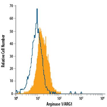
Detection of Arginase 1/ARG1 in HepG2 Human Cell Line by Flow Cytometry.
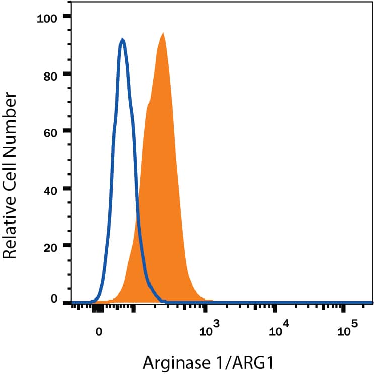
HepG2 human hepatocellular carcinoma cell line was stained with Sheep Anti-Human/Mouse Arginase 1/ARG1 APC-conjugated Antigen Affinity-purified Polyclonal Antibody (Catalog # IC5868A, filled histogram) or isotype control antibody (Catalog # IC016A, open histogram). To facilitate intracellular staining, cells were fixed with Flow Cytometry Fixation Buffer (Catalog # FC004) and permeabilized with Flow Cytometry Permeabilization/Wash Buffer I (Catalog # FC005). View our protocol for Staining Intracellular Molecules.Detection of Arginase 1/ARG1 in Hepa 1-6 Mouse Cell Line by Flow Cytometry.
Hepa 1-6 mouse hepatoma cell line was stained with Sheep Anti-Human/Mouse Arginase 1/ARG1 APC-conjugated Antigen Affinity-purified Polyclonal Antibody (Catalog # IC5868A, filled histogram) or isotype control antibody (Catalog # IC016A, open histogram). To facilitate intracellular staining, cells were fixed with Flow Cytometry Fixation Buffer (Catalog # FC004) and permeabilized with Flow Cytometry Permeabilization/Wash Buffer I (Catalog # FC005). View our protocol for Staining Intracellular Molecules.Detection of Mouse Arginase 1/ARG1/liver Arginase by Flow Cytometry
ILC2 transfers to apoE−/− mice increase peritoneal B1 cells, eosinophils and AAMs. Flow cytometry gating strategies of B1 cells identified as CD19+B220lowCD11b+IgM+ (a), eosinophils as CD45+SiglecF+CD11b+ cells (d) and AAMs as CD45+CD11b+F480+Arg1+ cells (i) of apoE−/− mice for both control and ILC2 groups respectively as indicated. Percentages and numbers of peritoneal B1 cells (b, c), eosinophils (e, f) and AAMs (j, k). CD11c+ expression from gated eosinophils and respective cell numbers of activated eosinophils identified as CD45+SiglecF+CD11b+CD11c+cells (g, h). Mann Whitney U test, **P < 0.01, ***P < 0.001, ****P < 0.0001. Each data point represents one mouse Image collected and cropped by CiteAb from the following publication (https://pubmed.ncbi.nlm.nih.gov/31823769), licensed under a CC-BY license. Not internally tested by R&D Systems.Applications for Human/Mouse Arginase 1/ARG1 APC-conjugated Antibody
Intracellular Staining by Flow Cytometry
Sample: HepG2 human hepatocellular carcinoma cell line and Hepa 1-6 mouse hepatoma cell line fixed with Flow Cytometry Fixation Buffer (Catalog # FC004) and permeabilized with Flow Cytometry Permeabilization/Wash Buffer I (Catalog # FC005).
Reviewed Applications
Read 1 review rated 4 using IC5868A in the following applications:
Formulation, Preparation, and Storage
Purification
Formulation
Shipping
Stability & Storage
- 12 months from date of receipt, 2 to 8 °C as supplied.
Background: Arginase 1/ARG1
Arginase 1 (ARG1) is a 35‑40 kDa member of the arginase family of enzymes. It is expressed in multiple cell types, including erythrocytes, hepatocytes, neutrophils, smooth muscle and macrophages. ARG1 demonstrates two distinct functions: in the hepatocyte cytoplasm, it catalyzes the conversion of arginine to ornithine and urea, while in multiple cells, it degrades arginine, thus indirectly downregulating NO synthase (NOS) activity by depriving this enzyme of its substrate. Human AGR1 is 322 amino acids (aa) in length. Its enzyme region comprises aa 9‑309 and contains two Mn atoms. ARG1 is modestly active as a monomer, but highly active as a 105 kDa homotrimer. Trimerization is promoted by nitrosylation of Cys303, creating a regulatory feedback loop with NOS. There are two isoform variants, one that shows an eight aa insertion after Gln43, and another that shows a deletion of aa 204‑289. Full-length human ARG1 shares 87% aa identity with mouse and rat ARG1.
Long Name
Alternate Names
Gene Symbol
UniProt
Additional Arginase 1/ARG1 Products
Product Documents for Human/Mouse Arginase 1/ARG1 APC-conjugated Antibody
Product Specific Notices for Human/Mouse Arginase 1/ARG1 APC-conjugated Antibody
For research use only


