Human/Mouse BCAP Antibody
R&D Systems, part of Bio-Techne | Catalog # AF4857

Key Product Details
Species Reactivity
Validated:
Cited:
Applications
Validated:
Cited:
Label
Antibody Source
Product Specifications
Immunogen
Met462-Leu650
Accession # Q6ZUJ8
Specificity
Clonality
Host
Isotype
Scientific Data Images for Human/Mouse BCAP Antibody
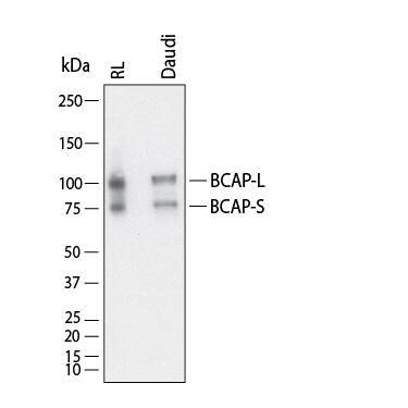
Detection of Human BCAP by Western Blot.
Western Blot shows lysates of RL human non-Hodgkin's lymphoma B cell line and Daudi human Burkitt's lymphoma cell line. PVDF membrane was probed with 1 µg/ml of Goat Anti-Human/Mouse BCAP Antigen Affinity-purified Polyclonal Antibody (Catalog # AF4857) followed by HRP-conjugated Anti-Goat IgG Secondary Antibody (Catalog # HAF017). Specific bands were detected for BCAP at approximately 80 kDa and 100 kDa (as indicated). This experiment was conducted under reducing conditions and using Western Blot Buffer Group 1.Detection of Human BCAP/PIK3AP1 by Western Blot
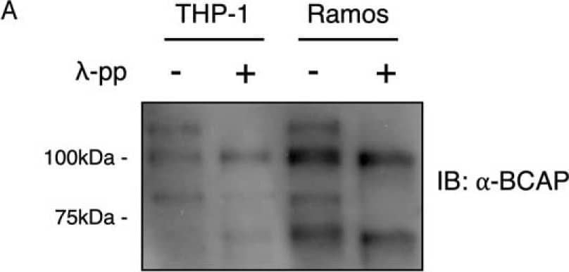
BCAP is hyperphosphorylated in B-cell, macrophages, and Expi293F cells.A, lysates from THP-1 and Ramos cells were dephosphorylated with lambda-phosphatase and immunoblotted for BCAP. B, His-Avi-tagged BCAP expressed in Expi293F cells was purified and dephosphorylated with lambda-phosphatase before immunostaining for tyrosine and serine phosphorylation. C, phosphorylation sites of BCAP expressed in Expi293F cells were determined by phosphopeptide mapping. BCAP was digested with trypsin, chymotrypsin, Asp-N, and Glu-C prior to MS. Image collected and cropped by CiteAb from the following open publication (https://pubmed.ncbi.nlm.nih.gov/31527084), licensed under a CC-BY license. Not internally tested by R&D Systems.Applications for Human/Mouse BCAP Antibody
Western Blot
Sample: RL human non-Hodgkin's lymphoma B cell line and Daudi human Burkitt's lymphoma cell line
Formulation, Preparation, and Storage
Purification
Reconstitution
Formulation
*Small pack size (-SP) is supplied either lyophilized or as a 0.2 µm filtered solution in PBS.
Shipping
Stability & Storage
- 12 months from date of receipt, -20 to -70 °C as supplied.
- 1 month, 2 to 8 °C under sterile conditions after reconstitution.
- 6 months, -20 to -70 °C under sterile conditions after reconstitution.
Background: BCAP
BCAP (B-cell adaptor for phosphoinositide 3-kinase (PI3K)) participates in linking the B cell antigen receptor (BCR) with the PI3K pathway. Tyrosine phosphorylation of BCAP by BCR-associated protein tyrosine kinases, such as Syk and Btk, generates binding sites for the p85 subunit of PI3K, resulting in activation of the PI3K pathway. Human and mouse BCAP contain 3 YXXM SH2 binding motifs for interaction with PI3K.
Long Name
Alternate Names
Gene Symbol
UniProt
Additional BCAP Products
Product Documents for Human/Mouse BCAP Antibody
Product Specific Notices for Human/Mouse BCAP Antibody
For research use only

