Human/Mouse CBFB Antibody
R&D Systems, part of Bio-Techne | Catalog # AF7349

Key Product Details
Species Reactivity
Applications
Label
Antibody Source
Product Specifications
Immunogen
Pro2-Glu165
Accession # Q13951
Specificity
Clonality
Host
Isotype
Scientific Data Images for Human/Mouse CBFB Antibody
Detection of Human and Mouse CBFB by Western Blot.
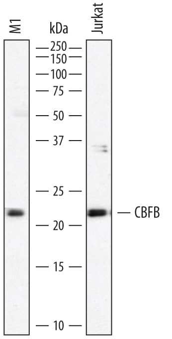
Western blot shows lysates of M1 mouse myeloid leukemia cell line and Jurkat human acute T cell leukemia cell line. PVDF membrane was probed with 1 µg/mL of Sheep Anti-Human CBFB Antigen Affinity-purified Polyclonal Antibody (Catalog # AF7349) followed by HRP-conjugated Anti-Sheep IgG Secondary Antibody (Catalog # HAF016). A specific band was detected for CBFB at approximately 22 kDa (as indicated). This experiment was conducted under reducing conditions and using Immunoblot Buffer Group 1.CBFB in HUVEC Human Cells.
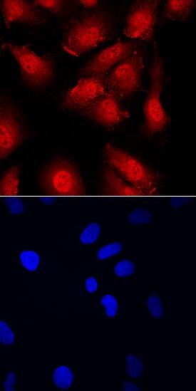
CBFB was detected in immersion fixed HUVEC human umbilical vein endothelial cells using Sheep Anti-Human/Mouse CBFB Antigen Affinity-purified Polyclonal Antibody (Catalog # AF7349) at 10 µg/mL for 3 hours at room temperature. Cells were stained using the NorthernLights™ 557-conjugated Anti-Sheep IgG Secondary Antibody (red, upper panel; Catalog # NL010) and counterstained with DAPI (blue, lower panel). Specific staining was localized to cytoplasm and nuclei. View our protocol for Fluorescent ICC Staining of Cells on Coverslips.Applications for Human/Mouse CBFB Antibody
Immunocytochemistry
Sample: Immersion fixed human umbilical vein endothelial cells (HUVECs)
Western Blot
Formulation, Preparation, and Storage
Purification
Reconstitution
Formulation
Shipping
Stability & Storage
- 12 months from date of receipt, -20 to -70 °C as supplied.
- 1 month, 2 to 8 °C under sterile conditions after reconstitution.
- 6 months, -20 to -70 °C under sterile conditions after reconstitution.
Background: CBFB
CBFB (Core binding Factor beta; also PEA2 beta and PEBP2 beta) is a widely-expressed 21-24 kDa member of the CBFB family of proteins. It forms dimeric transcriptional complexes with multiple molecules, including CBFB, RUNX1 and RUNX2. Although it does not bind DNA, it potentiates the binding of its partners to DNA. Human CBFB is 182 amino acids (aa) in length. It contains multiple alpha-helices and beta-strands. CBFB has at least two splice variants. One is approximately 23 kDa in size and possesses a 22 aa substitution for aa 166-182. A second is 16 kDa in size and contains a 22 aa substitution for aa 134-182. And a third shows a potential deletion of aa 56-94 coupled to the above 22 substitution for aa 166-182. CBFB is known to form a 68-70 kDa fusion protein with smooth muscle myosin heavy chain in AML. The CBFB contribution to the N-terminus of this fusion protein usually involves aa 1-165. Over aa 1-165, human CBFB shares 98% aa sequence identity with mouse CBFB.
Long Name
Alternate Names
Gene Symbol
UniProt
Additional CBFB Products
Product Documents for Human/Mouse CBFB Antibody
Product Specific Notices for Human/Mouse CBFB Antibody
For research use only

