Human/Mouse cIAP Pan-specific Antibody
R&D Systems, part of Bio-Techne | Catalog # MAB3400

Key Product Details
Validated by
Biological Validation
Species Reactivity
Validated:
Human, Mouse
Cited:
Human, Mouse, Avian - Chicken
Applications
Validated:
Simple Western, Western Blot
Cited:
Flow Cytometry, Immunohistochemistry, Immunohistochemistry-Paraffin, Immunoprecipitation, Western Blot
Label
Unconjugated
Antibody Source
Monoclonal Mouse IgG2A Clone # 315301
Product Specifications
Immunogen
E. coli-derived recombinant human cIAP-2
Asn2-Ser604
Accession # Q13489
Asn2-Ser604
Accession # Q13489
Specificity
Detects human and mouse cIAP-1 and cIAP-2 in Western blots.
Clonality
Monoclonal
Host
Mouse
Isotype
IgG2A
Scientific Data Images for Human/Mouse cIAP Pan-specific Antibody
Detection of Human/Mouse cIAP by Western Blot.
Western blot shows lysates of HEK293 human embryonic kidney cell line transfected with human cIAP-1 or human cIAP-2. PVDF membrane was probed with 0.5 µg/mL Mouse Anti-Human/Mouse cIAP Pan-specific Monoclonal Antibody (Catalog # MAB3400) followed by HRP-conjugated Anti-Mouse IgG Secondary Antibody (Catalog # HAF007). For additional reference, lysates of Raji human Burkitt's lymphoma cell line, Jurkat human acute T cell leukemia cell line, and SW3T3 mouse contact inhibited fibroblast cell line were included. Specific bands for cIAP-1 and cIAP-2 were detected at approximately 80 kDa (as indicated). This experiment was conducted under reducing conditions and using Immunoblot Buffer Group 2.Detection of Human cIAP by Simple WesternTM.
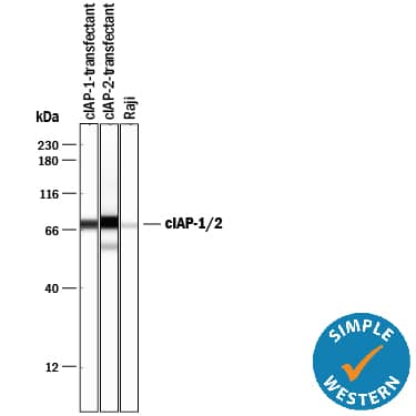
Simple Western lane view shows lysates of HEK293 human embryonic kidney cell line transfected with either cIAP-1 or cIAP-2 and Raji human Burkitt's lymphoma cell line, loaded at 0.2 mg/mL. A specific band was detected for cIAP-1 and cIAP-2 at approximately 73 kDa (as indicated) using 10 µg/mL of Mouse Anti-Human/Mouse cIAP Pan-specific Monoclonal Antibody (Catalog # MAB3400). This experiment was conducted under reducing conditions and using the 12-230 kDa separation system.Detection of Human cIAP (pan) by Western Blot
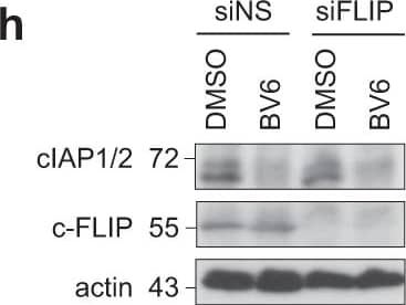
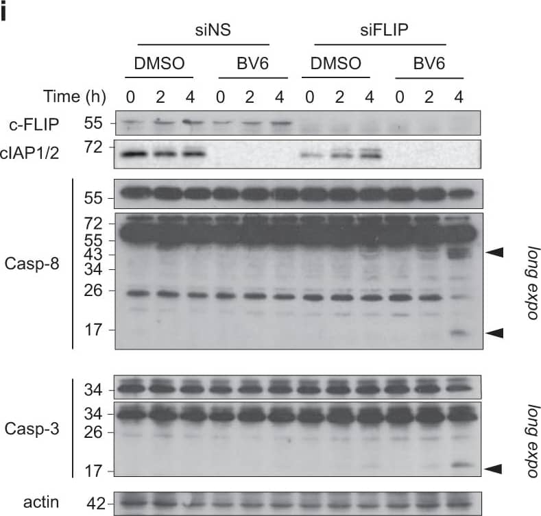
cIAPs and c-FLIP exert an efficient double brake against TLR3-mediated apoptosis in normal bronchial epithelial cells.a Primary human bronchial epithelial cells (HBEC) were exposed to Poly(I:C) (100 μg/ml) for 24 h, and the secretion of chemokines/cytokines were determined. Error bars represent S.E.M. of two independent experiments. b Primary HBEC were exposed to Poly(I:C) (100 μg/ml) for 6 and 24 h (left panel), or to cisplatin for 24 h (right panel), and the percentage of Annexin V+ cells was determined by flow cytometry. Error bars represent S.E.M. of three (left panel) and two (right panel) independent experiments. c Human immortalized bronchial epithelial HBEC3-KT cells were treated with Poly(I:C) (100 μg/ml) for 6 h and 24 h (left panel), or to cisplatin (100 μM) for 24 h (right panel), and the percentage of Annexin V+ cells was determined. Error bars represent S.E.M. of four (left panel) and three (right panel) independent experiments. **P < 0.01. d HBEC3-KT cells were transfected with a control non-silencing siRNA (siNS) or targeting TLR3 (siTLR3), exposed to Poly(I:C) (100 μg/ml) for 24 h, and the secretion of chemokines/cytokines was determined by ELISA. Error bars represent S.E.M. of three independent experiments. *P < 0.05. e HBEC3-KT cells were transfected as in d, lysed, and immunoblotted as indicated. f The indicated cells were lysed and immunoblotted for TLR3 and actin. Representative images of three independent experiments. g HBEC3-KT cells transfected with a control siRNA (siNS) or targeting c-FLIP (siFLIP), and were treated for 1 h with BV6 (5 μM). The cells were then exposed for 6 h to increasing doses of Poly(I:C), and the percentage of Annexin V+ cells was determined. Error bars represent S.E.M. of four independent experiments. *P < 0.05 and **P < 0.01. h HBEC3-KT cells were treated as in f, lysed, and immunoblotted as indicated. i HBEC3-KT transfected with siNS or siFLIP were then exposed to Poly(I:C) (100 μg/ml) for the indicated times in presence or absence of BV6 (5 μM). The cells were then lysed and immunoblotted as indicated. Caspase cleavage products are indicated by arrowheads. Representative images of two independent experiments Image collected and cropped by CiteAb from the following publication (https://pubmed.ncbi.nlm.nih.gov/30158588), licensed under a CC-BY license. Not internally tested by R&D Systems.Applications for Human/Mouse cIAP Pan-specific Antibody
Application
Recommended Usage
Simple Western
10 µg/mL
Sample: HEK293 human embryonic kidney cell line transfected with either cIAP-1 or cIAP-2 and Raji human Burkitt's lymphoma cell line
Sample: HEK293 human embryonic kidney cell line transfected with either cIAP-1 or cIAP-2 and Raji human Burkitt's lymphoma cell line
Western Blot
0.5 µg/mL
Sample: HEK293 human embryonic kidney cell line transfected with human cIAP-1 or human cIAP-2, Raji human Burkitt's lymphoma cell line, Jurkat human acute T cell leukemia cell line, and SW3T3 mouse contact inhibited fibroblast cell line
Sample: HEK293 human embryonic kidney cell line transfected with human cIAP-1 or human cIAP-2, Raji human Burkitt's lymphoma cell line, Jurkat human acute T cell leukemia cell line, and SW3T3 mouse contact inhibited fibroblast cell line
Formulation, Preparation, and Storage
Purification
Protein A or G purified from hybridoma culture supernatant
Reconstitution
Reconstitute at 0.5 mg/mL in sterile PBS. For liquid material, refer to CoA for concentration.
Formulation
Lyophilized from a 0.2 μm filtered solution in PBS with Trehalose. *Small pack size (SP) is supplied either lyophilized or as a 0.2 µm filtered solution in PBS.
Shipping
Lyophilized product is shipped at ambient temperature. Liquid small pack size (-SP) is shipped with polar packs. Upon receipt, store immediately at the temperature recommended below.
Stability & Storage
Use a manual defrost freezer and avoid repeated freeze-thaw cycles.
- 12 months from date of receipt, -20 to -70 °C as supplied.
- 1 month, 2 to 8 °C under sterile conditions after reconstitution.
- 6 months, -20 to -70 °C under sterile conditions after reconstitution.
Background: cIAP (pan)
Additional cIAP (pan) Products
Product Documents for Human/Mouse cIAP Pan-specific Antibody
Product Specific Notices for Human/Mouse cIAP Pan-specific Antibody
For research use only
Loading...
Loading...
Loading...
Loading...


