Human/Mouse Cyclin D1/D2 Antibody
R&D Systems, part of Bio-Techne | Catalog # AF4196

Key Product Details
Species Reactivity
Validated:
Cited:
Applications
Validated:
Cited:
Label
Antibody Source
Product Specifications
Immunogen
Met1-Ile295
Accession # P24385
Specificity
Clonality
Host
Isotype
Scientific Data Images for Human/Mouse Cyclin D1/D2 Antibody
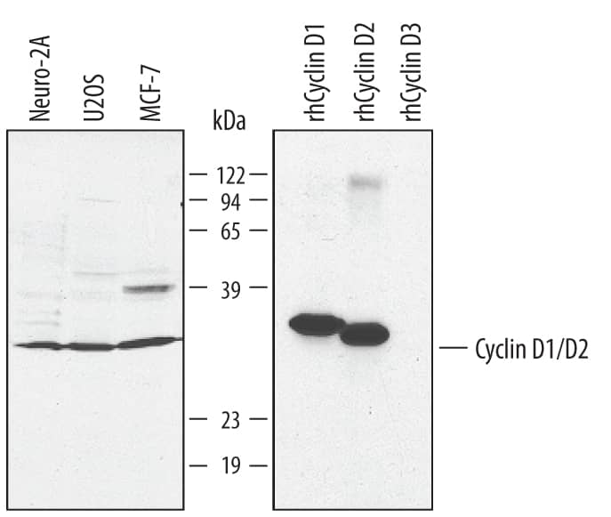
Detection of Human/Mouse Cyclin D1/D2 by Western Blot.
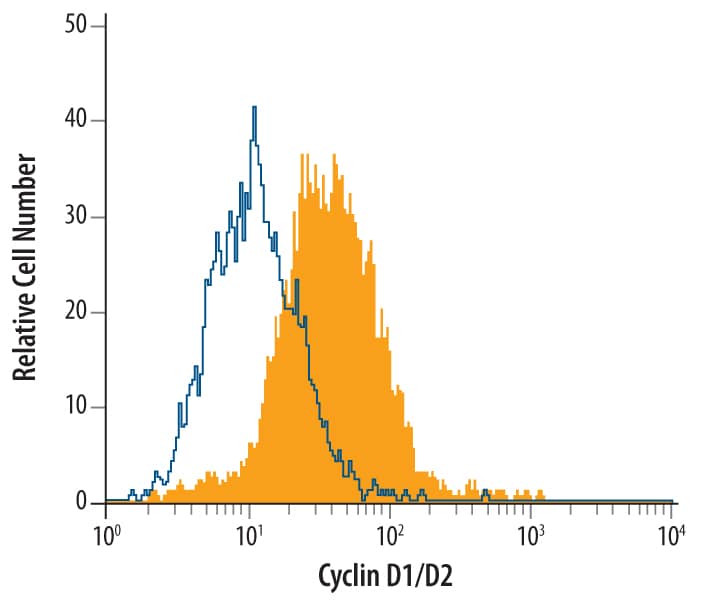
Western blot shows lysates of Neuro-2A mouse neuroblastoma cell line, U2OS human osteosarcoma cell line, and MCF-7 human breast cancer cell line. PVDF membrane was probed with 1 µg/mL Goat Anti-Human/Mouse Cyclin D1/D2 Cross-reactive Antigen Affinity-purified Polyclonal Antibody (Catalog # AF4196) followed by HRP-conjugated Anti-Goat IgG Secondary Antibody (Catalog # HAF017). For additional reference, recombinant human Cyclin D1, D2, and D3 (5 ng/lane) were included. A specific band for Cyclin D1 was detected at approximately 33 kDa (as indicated). This experiment was conducted under reducing conditions and using Immunoblot Buffer Group 1.Detection of Cyclin D1/D2 in MCF‑7 Human Cell Line by Flow Cytometry.
MCF-7 human breast cancer cell line was stained with Goat Anti-Human/Mouse Cross-reactive Antigen Affinity-purified Polyclonal Antibody (Catalog # AF4196, filled histogram) or isotype control AA175antibody (Catalog # AB-108-C, open histogram), followed by Phycoerythrin-conjugated Anti-Goat IgG Secondary Antibody (Catalog # F0107). To facilitate intracellular staining, cells were fixed with paraformaldeyde and permeabilized with saponin.Applications for Human/Mouse Cyclin D1/D2 Antibody
CyTOF-ready
Intracellular Staining by Flow Cytometry
Sample:
MCF‑7 human breast cancer cell line fixed with paraformaldeyde and permeabilized with saponin.
Western Blot
Sample: Neuro-2A mouse neuroblastoma cell line, U2OS human osteosarcoma cell line, and MCF-7 human breast cancer cell line
Formulation, Preparation, and Storage
Purification
Reconstitution
Formulation
Shipping
Stability & Storage
- 12 months from date of receipt, -20 to -70 °C as supplied.
- 1 month, 2 to 8 °C under sterile conditions after reconstitution.
- 6 months, -20 to -70 °C under sterile conditions after reconstitution.
Background: Cyclin D1/D2
The D-type cyclins (cyclins D1, D2, and D3) and their associated kinases, Cdk4 and Cdk6, play an important role in the progression from G0/G1 to S phase in the mammalian cell cycle. Cyclin D complexes containing Cdk4 or Cdk6 phosphorylate the retinoblastoma protein (pRb). pRb phosphorylation leads to the release of E2F transcription factors, which activate the expression of S-phase genes and thereby induce cell cycle progression. Cyclin D1, independent of Cdk4 activity, functions as a transcriptional modulator by regulating the activity of several transcription factors, such as estrogen receptor, Myb, and STAT3. Cyclin D1 has also been linked to the development and progression of several cancers including breast, bladder, esophagus, and lung. Overexpression of Cyclin D2 has been reported in gastric carcinoma, ovarian granulose cell tumors, and hematopoietic cell cancers, while in breast cancers, cyclin D2 expression is undetectable in 80% of the tumors.
Alternate Names
Gene Symbol
UniProt
Additional Cyclin D1/D2 Products
Product Documents for Human/Mouse Cyclin D1/D2 Antibody
Product Specific Notices for Human/Mouse Cyclin D1/D2 Antibody
For research use only

