Human/Mouse Desmocollin-1 Antibody
R&D Systems, part of Bio-Techne | Catalog # MAB7367

Conjugate
Catalog #
Key Product Details
Species Reactivity
Validated:
Human, Mouse
Cited:
Human, Mouse
Applications
Validated:
CyTOF-ready, Flow Cytometry, Immunocytochemistry, Immunohistochemistry, Western Blot
Cited:
Immunohistochemistry-Frozen, Western Blot
Label
Unconjugated
Antibody Source
Monoclonal Rat IgG2A Clone # 772906
Product Specifications
Immunogen
Mouse myeloma cell line NS0-derived recombinant mouse Desmocollin-1
Arg135-Lys691
Accession # P55849
Arg135-Lys691
Accession # P55849
Specificity
Detects mouse Desmocollin-1 in ELISAs and Western blots. In direct ELISAs, 100% cross-reactivity with
recombinant human Desmocollin-1 is observed, and no cross-reactivity with
recombinant mouse Desmocollin-2 or -3 is observed.
Clonality
Monoclonal
Host
Rat
Isotype
IgG2A
Scientific Data Images for Human/Mouse Desmocollin-1 Antibody
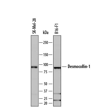
Detection of Human and Mouse Desmocollin‑1 by Western Blot.
Western blot shows lysates of SK-Mel-28 human malignant melanoma cell line and B16-F1 mouse melanoma cell line. PVDF membrane was probed with 0.1 µg/mL of Rat Anti-Human/Mouse Desmocollin-1 Monoclonal Antibody (Catalog # MAB7367) followed by HRP-conjugated Anti-Rat IgG Secondary Antibody (Catalog # HAF005). A specific band was detected for Desmocollin-1 at approximately 98 kDa (as indicated). This experiment was conducted under reducing conditions and using Immunoblot Buffer Group 1.Detection of Desmocollin-1 in B16‑F1 Mouse Cell Line by Flow Cytometry.
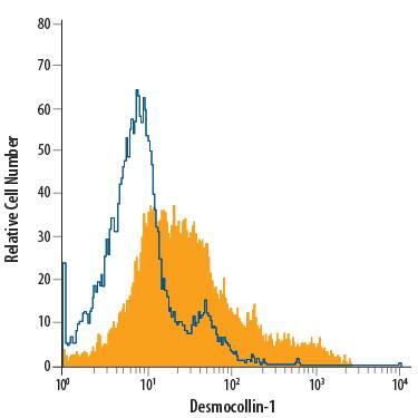
B16-F1 mouse melanoma cell line was stained with Rat Anti-Human/Mouse Desmocollin-1 Monoclonal Antibody (Catalog # MAB7367, filled histogram) or isotype control antibody (Catalog # MAB006, open histogram), followed by Allophycocyanin-conjugated Anti-Rat IgG Secondary Antibody (Catalog # F0113).Detection of Desmocollin‑1 in A549 Human Cell Line by Flow Cytometry.
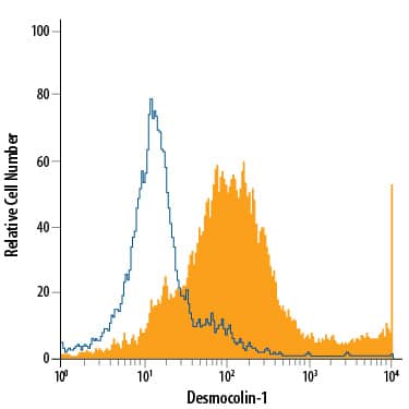
A549 human lung carcinoma cell line was stained with Rat Anti-Human/Mouse Desmocollin-1 Monoclonal Antibody (Catalog # MAB7367, filled histogram) or isotype control antibody (Catalog # MAB006, open histogram), followed by Allophycocyanin-conjugated Anti-Rat IgG Secondary Antibody (Catalog # F0113).Applications for Human/Mouse Desmocollin-1 Antibody
Application
Recommended Usage
CyTOF-ready
Ready to be labeled using established conjugation methods. No BSA or other carrier proteins that could interfere with conjugation.
Flow Cytometry
0.25 µg/106 cells
Sample: B16‑F1 mouse melanoma cell line and A549 human lung carcinoma cell line
Sample: B16‑F1 mouse melanoma cell line and A549 human lung carcinoma cell line
Immunocytochemistry
8-25 µg/mL
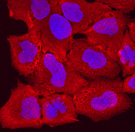
Sample: Immersion fixed A549 human lung carcinoma cell line
Sample: Immersion fixed A549 human lung carcinoma cell line
Immunohistochemistry
1-25 µg/mL
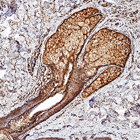
Sample: Perfusion fixed frozen sections of mouse skin
Sample: Perfusion fixed frozen sections of mouse skin
Western Blot
0.1 µg/mL
Sample: SK‑Mel‑28 human malignant melanoma cell line and B16‑F1 mouse melanoma cell line
Sample: SK‑Mel‑28 human malignant melanoma cell line and B16‑F1 mouse melanoma cell line
Reviewed Applications
Read 1 review rated 5 using MAB7367 in the following applications:
Formulation, Preparation, and Storage
Purification
Protein A or G purified from hybridoma culture supernatant
Reconstitution
Sterile PBS to a final concentration of 0.5 mg/mL. For liquid material, refer to CoA for concentration.
Formulation
Lyophilized from a 0.2 μm filtered solution in PBS with Trehalose. *Small pack size (SP) is supplied either lyophilized or as a 0.2 µm filtered solution in PBS.
Shipping
Lyophilized product is shipped at ambient temperature. Liquid small pack size (-SP) is shipped with polar packs. Upon receipt, store immediately at the temperature recommended below.
Stability & Storage
Use a manual defrost freezer and avoid repeated freeze-thaw cycles.
- 12 months from date of receipt, -20 to -70 °C as supplied.
- 1 month, 2 to 8 °C under sterile conditions after reconstitution.
- 6 months, -20 to -70 °C under sterile conditions after reconstitution.
Background: Desmocollin-1
DSC‑1 expression is induced by DSG-1. It serves as a component of desmosomes, forming a linkage that unites adjacent cells with cytoplasmic intermediate filaments. In particular, homodimeric DSC-1 may form heterotypic interactions with DSG-1 in-trans, and bind to the cytoskeleton intracellularly via plakophilin-1. Mature mouse DSC-1 is a 760 amino acid (aa) type I transmembrane glycoprotein (aa 135-691). The mature molecule contains a 557 aa extracellular region with five cadherin domains (aa 135-682), and a 172 aa cytoplasmic domain. There is one splice variant that shows an 11 aa substitution for aa 822-886. Over aa 135-691, mouse DSC‑1 shares 82% aa sequence identity with human DSC-1.
Alternate Names
CDHF1, Desmocollin1, DG2/DG3, DSC1
Gene Symbol
DSC1
UniProt
Additional Desmocollin-1 Products
Product Documents for Human/Mouse Desmocollin-1 Antibody
Product Specific Notices for Human/Mouse Desmocollin-1 Antibody
For research use only
Loading...
Loading...
Loading...
Loading...
Loading...
