Human/Mouse FLIP Antibody
R&D Systems, part of Bio-Techne | Catalog # AF821

Key Product Details
Species Reactivity
Validated:
Human, Mouse
Cited:
Human, Mouse, Porcine
Applications
Validated:
Western Blot
Cited:
Flow Cytometry, Immunocytochemistry, Immunohistochemistry, Western Blot
Label
Unconjugated
Antibody Source
Polyclonal Rabbit IgG
Product Specifications
Immunogen
KLH-coupled mouse FLIP synthetic peptide
LKQGRRRPLVDLHVELMDKC
LKQGRRRPLVDLHVELMDKC
Specificity
Detects human and mouse FLIP in Western blots.
Clonality
Polyclonal
Host
Rabbit
Isotype
IgG
Scientific Data Images for Human/Mouse FLIP Antibody
Detection of Human/Mouse FLIP by Western Blot.
Western blot shows lysates of WS-1 human fetal skin fibroblast cell line and L-929 mouse fibroblast cell line. PVDF membrane was probed with 1 µg/mL of Rabbit Anti-Human/Mouse FLIP Antigen Affinity-purified Polyclonal Antibody (Catalog # AF821) followed by HRP-conjugated Anti-Rabbit IgG Secondary Antibody (Catalog # HAF008). A specific band was detected for FLIP at approximately 56 kDa (as indicated). This experiment was conducted under reducing conditions and using Immunoblot Buffer Group 2.Detection of Mouse FLIP by Western Blot
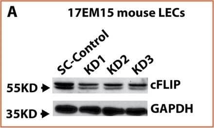
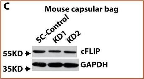
TNF alpha and CHX trigger an apoptotic response in cFlip knockdown 17EM15 cells and ex vivo cultured mouse lens capsular bag by activating caspase-8, caspase-9, and caspase-3.A cFlip knocking down in 17EM15 mouse LECs determined by immunoblot-assay. B The semi-quantitative densitometric measurement of 17EM15 cFlip knocking down from immunoblot-assay. C cFlip knocking down in ex vivo cultured mouse lens capsular bags. D The semi-quantitative densitometric measurement of cFlip knocking down mouse lens capsular bags from immunoblot-assay. E Pro-caspase-8, cleaved caspase-8, pro-caspase-3, and cleaved caspase-3 in cFlip knockdown and scrambled shRNA control (SC) 17EM15 cells with and without 30 ng/ml TNF alpha and 10μg/ml CHX stimulation for 7 h. F Pro-caspase-8, cleaved caspase-8, pro-caspase-3, and cleaved caspase-3 in cFlip knockdown and scrambled shRNA control (SC) ex vivo cultured mouse lens capsular bags with and without 60 ng/ml TNF alpha and 10 μg/ml CHX treatment for 24 h. The cFlip KD 17EM15 treated cell lysate was used as a positive control. Only 1/10th amount of protein relative to capsular bag lysate was loaded. One-way ANOVA was used to compare between groups, and only p < 0.05 is considered significant. *<0.05, **<0.01, ***<0.001, ****<0.0001. Image collected and cropped by CiteAb from the following open publication (https://pubmed.ncbi.nlm.nih.gov/33837174), licensed under a CC-BY license. Not internally tested by R&D Systems.Detection of Mouse FLIP by Western Blot
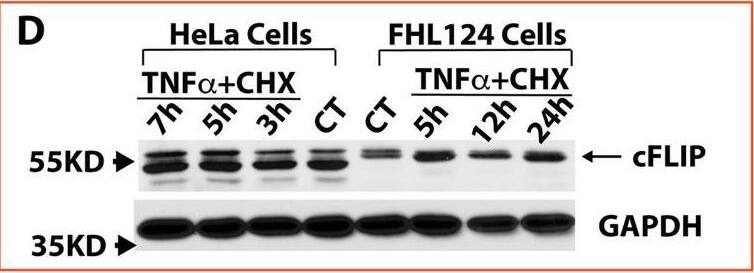
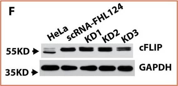
cFLIP is highly expressed in FHL124 cells and also significantly upregulated after TNF alpha stimulation.A Anti-apoptotic protein expression in FHL124 and HeLa cell with and without TNF alpha and TNF alpha plus CHX stimulation. B Relative cFLIP mRNA expression in FHL124 and HeLa cells with and without TNF alpha and CHX stimulation. FHL 124 cells demonstrated a more robust response to TNF alpha and CHX stimulation compared to HeLa cells. C The endogenous cFLIP, Caspase-8, caspase-9, and caspase-3 mRNA levels in FHL124 cells vs. HeLa cells. A much higher cFLIP mRNA expression was seen in FHL124 cells compared to HeLa cells. D,E cFLIP protein expression after TNF alpha and CHX stimulation in FHL124 and HeLa cells. cFLIP protein level was increased around 3-fold in FHL124 cells at 5 h after TNF alpha and CHX stimulation, while only a mild increase was seen in HeLa cells after stimulation. F,G FHL124 cells had 6-fold endogenous cFLIP protein levels vs. HeLa cells. KD3 shRNA was able to knock down 50% of cFLIP expression in FHL124 cells. Each assay was repeated at least three times. One-way ANOVA with Tukey’s Honest post-hoc analysis was used to compare between groups, and only p < 0.05 is considered significant. *<0.05, **<0.01, ***<0.001, ****<0.0001. Image collected and cropped by CiteAb from the following open publication (https://pubmed.ncbi.nlm.nih.gov/33837174), licensed under a CC-BY license. Not internally tested by R&D Systems.Applications for Human/Mouse FLIP Antibody
Application
Recommended Usage
Western Blot
1 µg/mL
Sample: WS-1 human fetal skin fibroblast cell line and L-929 mouse fibroblast cell line
Sample: WS-1 human fetal skin fibroblast cell line and L-929 mouse fibroblast cell line
Formulation, Preparation, and Storage
Purification
Antigen Affinity-purified
Reconstitution
Reconstitute at 0.2 mg/mL in sterile PBS. For liquid material, refer to CoA for concentration.
Formulation
Lyophilized from a 0.2 μm filtered solution in PBS with Trehalose. *Small pack size (SP) is supplied either lyophilized or as a 0.2 µm filtered solution in PBS.
Shipping
Lyophilized product is shipped at ambient temperature. Liquid small pack size (-SP) is shipped with polar packs. Upon receipt, store immediately at the temperature recommended below.
Stability & Storage
Use a manual defrost freezer and avoid repeated freeze-thaw cycles.
- 12 months from date of receipt, -20 to -70 °C as supplied.
- 1 month, 2 to 8 °C under sterile conditions after reconstitution.
- 6 months, -20 to -70 °C under sterile conditions after reconstitution.
Background: FLIP
Long Name
FLICE Inhibitory Proteins
Alternate Names
CASPER, CFLAR, FLAME-1, I-FLICE
Gene Symbol
CFLAR
Additional FLIP Products
Product Documents for Human/Mouse FLIP Antibody
Product Specific Notices for Human/Mouse FLIP Antibody
For research use only
Loading...
Loading...
Loading...
Loading...


