Human/Mouse Gastrokine 1 Antibody
R&D Systems, part of Bio-Techne | Catalog # AF7287

Key Product Details
Species Reactivity
Validated:
Cited:
Applications
Validated:
Cited:
Label
Antibody Source
Product Specifications
Immunogen
Tyr38-Tyr201
Accession # Q9CR36
Specificity
Clonality
Host
Isotype
Scientific Data Images for Human/Mouse Gastrokine 1 Antibody
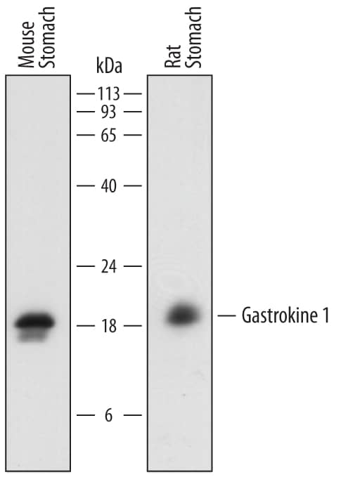
Detection of Mouse and Rat Gastrokine 1 by Western Blot.
Western blot shows lysates of mouse stomach tissue and rat stomach tissue. PVDF membrane was probed with 0.1 µg/mL of Sheep Anti-Human/Mouse Gastrokine 1 Antigen Affinity-purified Polyclonal Antibody (Catalog # AF7287) followed by HRP-conjugated Anti-Sheep IgG Secondary Antibody (Catalog # HAF016). A specific band was detected for Gastrokine 1 at approximately 18-20 kDa (as indicated). This experiment was conducted under reducing conditions and using Immunoblot Buffer Group 1.Gastrokine 1 in Mouse Stomach.
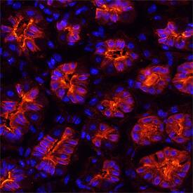
Gastrokine 1 was detected in perfusion fixed frozen sections of mouse stomach using Sheep Anti-Mouse Gastrokine 1 Antigen Affinity-purified Polyclonal Antibody (Catalog # AF7287) at 15 µg/mL overnight at 4 °C. Tissue was stained using the NorthernLights™ 557-conjugated Anti-Sheep IgG Secondary Antibody (red; Catalog # NL010) and counterstained with DAPI (blue). Specific staining was localized to islets. View our protocol for Fluorescent IHC Staining of Frozen Tissue Sections.Gastrokine 1 in Human Stomach.
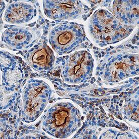
Gastrokine 1 was detected in immersion fixed paraffin-embedded sections of human stomach using Sheep Anti-Mouse Gastrokine 1 Antigen Affinity-purified Polyclonal Antibody (Catalog # AF7287) at 10 µg/mL overnight at 4 °C. Before incubation with the primary antibody, tissue was subjected to heat-induced epitope retrieval using Antigen Retrieval Reagent-Basic (Catalog # CTS013). Tissue was stained using the Anti-Sheep HRP-DAB Cell & Tissue Staining Kit (brown; Catalog # CTS019) and counterstained with hematoxylin (blue). Specific staining was localized to islets. View our protocol for Chromogenic IHC Staining of Paraffin-embedded Tissue Sections.Applications for Human/Mouse Gastrokine 1 Antibody
Immunohistochemistry
Sample: Immersion fixed paraffin-embedded sections of human stomach, and perfusion fixed frozen sections of mouse stomach
Western Blot
Sample: Mouse stomach tissue and rat stomach tissue
Formulation, Preparation, and Storage
Purification
Reconstitution
Formulation
Shipping
Stability & Storage
- 12 months from date of receipt, -20 to -70 °C as supplied.
- 1 month, 2 to 8 °C under sterile conditions after reconstitution.
- 6 months, -20 to -70 °C under sterile conditions after reconstitution.
Background: Gastrokine 1
Gastrokine-1 (GKN1; also CA11 and AMP-18) is an 18-20 kDa member of the Gastrokine protein family. It has limited expression, being restricted to mucous secreting pyloric antrum epithelial cells. Gastrokine-1 appears to promote epithelial cell proliferation and migration, and induce the formation of tight junctions between epithelial cells. By contrast, gastrokine-1 induces Fas expression in tumor cells, resulting in apoptosis. Mature mouse gastrokine-1 is 166 amino acids (aa) in length (aa 36-201). Based on the SwissProt sequence, it possesses one BRICHOS domain (aa 71-165) that contains a mitogenic sequence (aa 113-131). There is one potential alternative start site at Met18. Over aa 38-201, mouse gastrokine-1 shares 65% and 92% aa sequence identity with human and rat gastrokine-1, respectively.
Long Name
Alternate Names
Gene Symbol
UniProt
Additional Gastrokine 1 Products
Product Documents for Human/Mouse Gastrokine 1 Antibody
Product Specific Notices for Human/Mouse Gastrokine 1 Antibody
For research use only


