Human/Mouse Kir2.1 Antibody
R&D Systems, part of Bio-Techne | Catalog # MAB9548
Recombinant Monoclonal Antibody.

Key Product Details
Species Reactivity
Validated:
Human, Mouse
Cited:
Human, Mouse
Applications
Validated:
Immunocytochemistry, Immunohistochemistry, Western Blot
Cited:
Western Blot
Label
Unconjugated
Antibody Source
Recombinant Monoclonal Rabbit IgG Clone # 2153C
Product Specifications
Immunogen
E. coli-derived recombinant human Kir2.1
Asn352-Ile427
Accession # P63252
Asn352-Ile427
Accession # P63252
Specificity
Detects human Kir2.1 in direct ELISAs. Detects human and mouse Kir2.1 in Immunohistochemistry.
Clonality
Monoclonal
Host
Rabbit
Isotype
IgG
Scientific Data Images for Human/Mouse Kir2.1 Antibody
Detection of Human Kir2.1 by Western Blot.
Western blot shows lysates of Human heart ventricle tissue and human lung tissue. PVDF membrane was probed with 3 µg/mL of Rabbit Anti-Human/Mouse Kir2.1 Monoclonal Antibody (Catalog # MAB9548) followed by HRP-conjugated Anti-Rabbit IgG Secondary Antibody (Catalog # HAF008). A specific band was detected for Kir2.1 at approximately 50-55 kDa (as indicated). This experiment was conducted under reducing conditions and using Immunoblot Buffer Group 1.Kir2.1 in Human Heart.
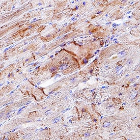
Kir2.1 was detected in immersion fixed paraffin-embedded sections of human heart using Rabbit Anti-Human/Mouse Kir2.1 Monoclonal Antibody (Catalog # MAB9548) at 3 µg/mL for 1 hour at room temperature followed by incubation with the Anti-Rabbit IgG VisUCyte™ HRP Polymer Antibody (Catalog # VC003). Tissue was stained using DAB (brown) and counterstained with hematoxylin (blue). Specific staining was localized to intercalated discs. View our protocol for IHC Staining with VisUCyte HRP Polymer Detection Reagents.Kir2.1 in Mouse Heart.
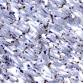
Kir2.1 was detected in perfusion fixed frozen sections of mouse heart using Rabbit Anti-Human/Mouse Kir2.1 Monoclonal Antibody (Catalog # MAB9548) at 3 µg/mL for 1 hour at room temperature followed by incubation with the Anti-Rabbit IgG VisUCyte™ HRP Polymer Antibody (Catalog # VC003). Tissue was stained using DAB (brown) and counterstained with hematoxylin (blue). Specific staining was localized to intercalated discs. View our protocol for IHC Staining with VisUCyte HRP Polymer Detection Reagents.Applications for Human/Mouse Kir2.1 Antibody
Application
Recommended Usage
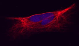
Immunocytochemistry
2-25 µg/mL
Sample: Immersion fixed human cardiomyocytes
Sample: Immersion fixed human cardiomyocytes
Immunohistochemistry
3-25 µg/mL
Sample: Immersion fixed paraffin-embedded sections of human heart and perfusion fixed frozen sections of mouse heart
Sample: Immersion fixed paraffin-embedded sections of human heart and perfusion fixed frozen sections of mouse heart
Western Blot
3 µg/mL
Sample: Human heart ventricle tissue and lung tissue
Sample: Human heart ventricle tissue and lung tissue
Reviewed Applications
Read 1 review rated 5 using MAB9548 in the following applications:
Formulation, Preparation, and Storage
Purification
Protein A or G purified from hybridoma culture supernatant
Reconstitution
Reconstitute at 0.5 mg/mL in sterile PBS. For liquid material, refer to CoA for concentration.
Formulation
Lyophilized from a 0.2 μm filtered solution in PBS with Trehalose. *Small pack size (SP) is supplied either lyophilized or as a 0.2 µm filtered solution in PBS.
Shipping
Lyophilized product is shipped at ambient temperature. Liquid small pack size (-SP) is shipped with polar packs. Upon receipt, store immediately at the temperature recommended below.
Stability & Storage
Use a manual defrost freezer and avoid repeated freeze-thaw cycles.
- 12 months from date of receipt, -20 to -70 °C as supplied.
- 1 month, 2 to 8 °C under sterile conditions after reconstitution.
- 6 months, -20 to -70 °C under sterile conditions after reconstitution.
Background: Kir2.1
Long Name
Inward Rectifier K(+) Channel Kir2.1
Alternate Names
ATFB9, HIRK1, IRK1, KCNJ2, LQT7, SQT3
Gene Symbol
KCNJ2
UniProt
Additional Kir2.1 Products
Product Documents for Human/Mouse Kir2.1 Antibody
Product Specific Notices for Human/Mouse Kir2.1 Antibody
For research use only
Loading...
Loading...
Loading...
Loading...


