Human/Mouse Metadherin Antibody
R&D Systems, part of Bio-Techne | Catalog # AF7180

Key Product Details
Species Reactivity
Applications
Label
Antibody Source
Product Specifications
Immunogen
Lys167-Ser297
Accession # Q80WJ7
Specificity
Clonality
Host
Isotype
Scientific Data Images for Human/Mouse Metadherin Antibody
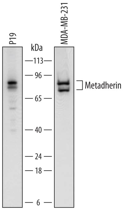
Detection of Human and Mouse Metadherin by Western Blot.
Western blot shows lysates of P19 mouse embryonal carcinoma cell line and MDA-MB-231 human breast cancer cell line. PVDF membrane was probed with 0.25 µg/mL of Sheep Anti-Human/Mouse Metadherin Antigen Affinity-purified Polyclonal Antibody (Catalog # AF7180) followed by HRP-conjugated Anti-Sheep IgG Secondary Antibody (Catalog # HAF016). Specific bands were detected for Metadherin at approximately 80 and 75 kDa (as indicated). This experiment was conducted under reducing conditions and using Immunoblot Buffer Group 1.Applications for Human/Mouse Metadherin Antibody
Western Blot
Sample: P19 mouse embryonal carcinoma cell line and MDA‑MB‑231 human breast cancer cell line
Formulation, Preparation, and Storage
Purification
Reconstitution
Formulation
Shipping
Stability & Storage
- 12 months from date of receipt, -20 to -70 °C as supplied.
- 1 month, 2 to 8 °C under sterile conditions after reconstitution.
- 6 months, -20 to -70 °C under sterile conditions after reconstitution.
Background: Metadherin
MTDH (Metadherin; also Lyric and Astrocyte-elevated gene-1/AEG1) is a unique molecule originally discovered to be upregulated in astrocytes following HIV infection. Although its predicted MW is 64 kDa, it runs anomalously at approximately 80 kDa in SDS-Page. It is widely expressed, and appears to be a component of the ER, nucleolus and inner nuclear membrane. MTDH is reported to promote Akt/PI3 kinase activity, and suppress apoptosis-associated FOXO3A transcription. Mouse MTDH is a type III (i.e.- a type I with no signal sequence) transmembrane protein 579 amino acids (aa) in length. It contains a luminal N-terminus (aa 1-48) with a 510 aa cytoplasmic C-terminus. There are no identifiable structural motifs, although two poly-Lys segments and two utilized Ser phosphorylation sites exist. Multiple bands at 80 kDa, 75 kDa, 50-55 kDa and 37 kDa are seen for MTDH on SDS-Page. They may reflect the presence of at least two potential isoform variants that show 1) a deletion of aa 422-457, and 2) an Ile substitution for aa 190-579. Over aa 167-297, mouse MTDH shares 98% and 97% aa sequence identity with rat and human MTDH, respectively.
Alternate Names
Gene Symbol
UniProt
Additional Metadherin Products
Product Documents for Human/Mouse Metadherin Antibody
Product Specific Notices for Human/Mouse Metadherin Antibody
For research use only
