Human/Mouse/Rat Akt1 Antibody
R&D Systems, part of Bio-Techne | Catalog # AF1775

Key Product Details
Species Reactivity
Validated:
Cited:
Applications
Validated:
Cited:
Label
Antibody Source
Product Specifications
Immunogen
Ser2-Ala480
Accession # P31749
Specificity
Clonality
Host
Isotype
Scientific Data Images for Human/Mouse/Rat Akt1 Antibody
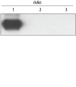
Detection of Human, Mouse, and Rat Akt1 by Western Blot.
Western blot shows lysates of HeLa human cervical epithelial carcinoma cell line, HepG2 human hepatocellular carcinoma cell line, TS1 mouse helper T cell line, and PC-12 rat adrenal pheochromocytoma cell line. PVDF membrane was probed with 0.1 µg/mL Rabbit Anti-Human/Mouse/Rat Akt1 Antigen Affinity-purified Polyclonal Antibody (Catalog # AF1775) followed by HRP-conjugated Anti-Rabbit IgG Secondary Antibody (Catalog # HAF008). A specific band for Akt1 was detected at approximately 60 kDa (as indicated). This experiment was conducted under reducing conditions and using Immunoblot Buffer Group 1.Detection of human Akt1 by Western Blot.
Western blot shows recombinant human Akt1, Akt2, and Akt3 (1 ng/lane). PVDF membrane was probed with 0.1 µg/mL Rabbit Anti-Human/Mouse/Rat Akt1 Antigen Affinity-purified Polyclonal Antibody (Catalog # AF1775) followed by HRP-conjugated Anti-Rabbit IgG Secondary Antibody (Catalog # HAF008). A specific band for Akt1 was detected at approximately 60 kDa (as indicated). This experiment was conducted under reducing conditions and using Immunoblot Buffer Group 1.Detection of Human Akt1 by Simple WesternTM.
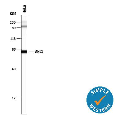
Simple Western lane view shows lysates of HeLa human cervical epithelial carcinoma cell line, loaded at 0.2 mg/mL. A specific band was detected for Akt1 at approximately 63 kDa (as indicated) using 1 µg/mL of Rabbit Anti-Human/Mouse/Rat Akt1 Antigen Affinity-purified Polyclonal Antibody (Catalog # AF1775). This experiment was conducted under reducing conditions and using the 12-230 kDa separation system. Non-specific interaction with the 230 kDa Simple Western standard may be seen with this antibody.Applications for Human/Mouse/Rat Akt1 Antibody
Simple Western
Sample: HeLa human cervical epithelial carcinoma cell line
Western Blot
Sample: HeLa human cervical epithelial carcinoma cell line, HepG2 human hepatocellular carcinoma cell line, TS1 mouse helper T cell line, and PC-12 rat adrenal pheochromocytoma cell line
Formulation, Preparation, and Storage
Purification
Reconstitution
Formulation
Shipping
Stability & Storage
- 12 months from date of receipt, -20 to -70 °C as supplied.
- 1 month, 2 to 8 °C under sterile conditions after reconstitution.
- 6 months, -20 to -70 °C under sterile conditions after reconstitution.
Background: Akt1
Akt, also known as protein kinase B (PKB), is a central kinase in such diverse cellular processes as glucose uptake, cell cycle progression, and apoptosis. Three highly homologous members define the Akt family: Akt1 (PKB alpha), Akt2 (PKB beta), and Akt3 (PKB gamma). Akt1 is the most ubiquitously expressed family member. All three Akts contain an amino-terminal pleckstrin homology domain, a central kinase domain, and a carboxyl-terminal regulatory domain.
Long Name
Alternate Names
Gene Symbol
UniProt
Additional Akt1 Products
Product Documents for Human/Mouse/Rat Akt1 Antibody
Product Specific Notices for Human/Mouse/Rat Akt1 Antibody
For research use only


