Human/Mouse/Rat Akt2 Antibody
R&D Systems, part of Bio-Techne | Catalog # AF23151

Key Product Details
Species Reactivity
Validated:
Cited:
Applications
Validated:
Cited:
Label
Antibody Source
Product Specifications
Immunogen
Accession # P31751
Specificity
Clonality
Host
Isotype
Scientific Data Images for Human/Mouse/Rat Akt2 Antibody
Detection of Human/Mouse/Rat Akt2 by Western Blot.
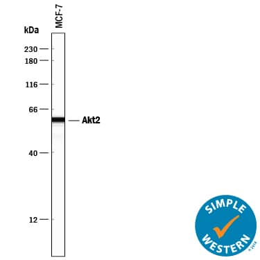
Western blot shows lysates of MCF-7 human breast cancer cell line, MDA-MB-468 human breast cancer cell line, and U937 human histiocytic lymphoma cell line. PVDF membrane was probed with 0.5 µg/mL Goat Anti-Human/Mouse/Rat Akt2 Antigen Affinity-purified Polyclonal Antibody (Catalog # AF23151) followed by HRP-conjugated Anti-Goat IgG Secondary Antibody (Catalog # HAF109). For additional reference, Recombinant Human Active Akt1 (Catalog # 1775-KS), recombinant human Akt2, and recombinant human Akt3 (5 ng/lane) were included. A specific band for Akt2 was detected at approximately 60 kDa (as indicated). This experiment was conducted under reducing conditions and using Immunoblot Buffer Group 1.Akt2 in Rat Testis.
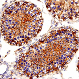
Akt2 was detected in perfusion fixed frozen sections of rat testis using Goat Anti-Human/Mouse/Rat Akt2 Antigen Affinity-purified Polyclonal Antibody (Catalog # AF23151) at 3 µg/mL for 1 hour at room temperature followed by incubation with the Anti-Goat IgG VisUCyte™ HRP Polymer Antibody (Catalog # VC004). Tissue was stained using DAB (brown) and counterstained with hematoxylin (blue). Specific staining was localized to cytoplasm and nuclei. View our protocol for IHC Staining with VisUCyte HRP Polymer Detection Reagents.Akt2 in Human Kidney Cancer Tissue.
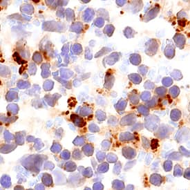
Akt2 was detected in immersion fixed paraffin-embedded sections of human kidney cancer tissue using 15 µg/mL Goat Anti-Human/Mouse/Rat Akt2 Antigen Affinity-purified Polyclonal Antibody (Catalog # AF23151) overnight at 4 °C. Tissue was stained with the Anti-Goat HRP-DAB Cell & Tissue Staining Kit (brown; Catalog # CTS008) and counterstained with hematoxylin (blue). Specific labeling was localized to epithelial cells in tubules. View our protocol for Chromogenic IHC Staining of Paraffin-embedded Tissue Sections.Applications for Human/Mouse/Rat Akt2 Antibody
Immunohistochemistry
Sample: Immersion fixed paraffin-embedded sections of human kidney cancer tissue, and perfusion fixed frozen sections of rat testis and mouse spleen
Simple Western
Sample: MCF‑7 human breast cancer cell line
Western Blot
Sample: MCF-7 human breast cancer cell line, MDA-MB-468 human breast cancer cell line, and U937 human histiocytic lymphoma cell line
Reviewed Applications
Read 1 review rated 4 using AF23151 in the following applications:
Formulation, Preparation, and Storage
Purification
Reconstitution
Formulation
Shipping
Stability & Storage
- 12 months from date of receipt, -20 to -70 °C as supplied.
- 1 month, 2 to 8 °C under sterile conditions after reconstitution.
- 6 months, -20 to -70 °C under sterile conditions after reconstitution.
Background: Akt2
The serine/threonine kinase Akt, also known as protein kinase B (PKB), is a central regulator of such diverse cellular processes as glucose uptake, cell cycle progression, and apoptosis. In mammals, three highly homologous members define the Akt family: Akt1 (PKB alpha), Akt2 (PKB beta), and Akt3 (PKB gamma). Akt2 is expressed predominantly in insulin target tissues such as liver, skeletal muscle, and fat. All three Akts contain an amino-terminal pleckstrin homology domain, a central kinase domain, and a carboxyl-terminal regulatory domain.
Long Name
Alternate Names
Gene Symbol
UniProt
Additional Akt2 Products
Product Documents for Human/Mouse/Rat Akt2 Antibody
Product Specific Notices for Human/Mouse/Rat Akt2 Antibody
For research use only


