Human/Mouse/Rat CHMP2B Antibody
R&D Systems, part of Bio-Techne | Catalog # AF7509

Key Product Details
Species Reactivity
Applications
Label
Antibody Source
Product Specifications
Immunogen
Ala2-Asp213
Accession # Q9UQN3
Specificity
Clonality
Host
Isotype
Scientific Data Images for Human/Mouse/Rat CHMP2B Antibody
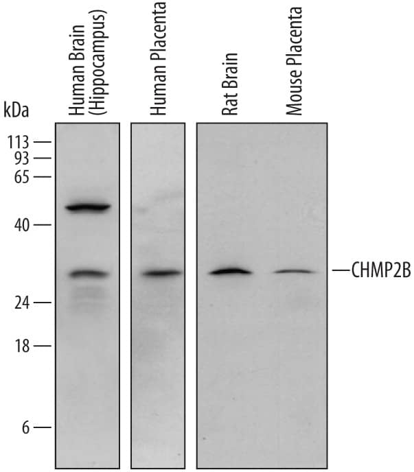
Detection of Human, Mouse, and Rat CHMP2B by Western Blot.
Western blot shows lysates of human brain (hippocampus) tissue, human placenta tissue, rat brain tissue, and mouse placenta tissue. PVDF membrane was probed with 1 µg/mL of Sheep Anti-Human/Mouse/Rat CHMP2B Antigen Affinity-purified Polyclonal Antibody (Catalog # AF7509) followed by HRP-conjugated Anti-Sheep IgG Secondary Antibody (Catalog # HAF016). A specific band was detected for CHMP2B at approximately 30 kDa (as indicated). This experiment was conducted under reducing conditions and using Immunoblot Buffer Group 1.CHMP2B in Human Brain.
CHMP2B was detected in immersion fixed paraffin-embedded sections of human brain (medulla) using Sheep Anti-Human/Mouse/Rat CHMP2B Antigen Affinity-purified Polyclonal Antibody (Catalog # AF7509) at 3 µg/mL overnight at 4 °C. Tissue was stained using the Anti-Sheep HRP-DAB Cell & Tissue Staining Kit (brown; Catalog # CTS019) and counterstained with hematoxylin (blue). Specific staining was localized to neuronal cell bodies. View our protocol for Fluorescent ICC Staining of Cells on Coverslips.Applications for Human/Mouse/Rat CHMP2B Antibody
Immunohistochemistry
Sample: Immersion fixed paraffin-embedded sections of human brain (medulla)
Western Blot
Sample: Human brain (hippocampus) tissue, human placenta tissue, rat brain tissue, and mouse placenta tissue
Formulation, Preparation, and Storage
Purification
Reconstitution
Formulation
Shipping
Stability & Storage
- 12 months from date of receipt, -20 to -70 °C as supplied.
- 1 month, 2 to 8 °C under sterile conditions after reconstitution.
- 6 months, -20 to -70 °C under sterile conditions after reconstitution.
Background: CHMP2B
CHMP2B (CHarged Multivescular body Protein 2B; also Chromatin-Modifying Protein 2B and Vps2-2) is a 35 kDa member of the SNF family of proteins. It is a cytosolic molecule that interacts with VPS4 and undergoes polymerization to form tubules that project plasma membrane outward, generating an environment conducive to membrane fission and budding. It also appears to participate in the formation of intraluminal vesicles associated with the lysosomal system. CHMP2B is found in striated muscle (heart and skeletal), and particularly in neurons of the CNS. Human CHMP2B is 213 amino acids (aa) in length. It contains one coiled-coil region (aa 25-55), an MIT (microtubule-interacting and trafficking) domain (aa 201-211) and a utilized phosphorylation site at Ser199. There is one alternative splice form that shows a deletion of aa 2-42. Full-length human CHMP2B shares 99% aa identity with mouse CHMP2B, but only 33% aa identity with human CHMP2A.
Long Name
Alternate Names
Gene Symbol
UniProt
Additional CHMP2B Products
Product Documents for Human/Mouse/Rat CHMP2B Antibody
Product Specific Notices for Human/Mouse/Rat CHMP2B Antibody
For research use only

