Human/Mouse/Rat DDX1 Antibody
R&D Systems, part of Bio-Techne | Catalog # MAB9615

Key Product Details
Species Reactivity
Applications
Label
Antibody Source
Product Specifications
Immunogen
Accession # Q92499
Specificity
Clonality
Host
Isotype
Scientific Data Images for Human/Mouse/Rat DDX1 Antibody
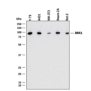
Detection of Human, Mouse, and Rat DDX1 by Western Blot.
Western blot shows lysates of Y-79 human retinoblastoma cell line, A431 human epithelial carcinoma cell line, NIH-3T3 mouse embryonic fibroblast cell line, Neuro-2A mouse neuroblastoma cell line, and Rat-2 rat embryonic fibroblast cell line. PVDF membrane was probed with 0.5 µg/mL of Rabbit Anti-Human/Mouse/Rat DDX1 Monoclonal Antibody (Catalog # MAB9615) followed by HRP-conjugated Anti-Rabbit IgG Secondary Antibody (Catalog # HAF008). A specific band was detected for DDX1 at approximately 85 kDa (as indicated). This experiment was conducted under reducing conditions and using Immunoblot Buffer Group 1.DDX1 in HeLa Human Cell Line.
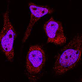
DDX1 was detected in immersion fixed HeLa human cervical epithelial carcinoma cell line using Rabbit Anti-Human/Mouse/Rat DDX1 Monoclonal Antibody (Catalog # MAB9615) at 1 µg/mL for 3 hours at room temperature. Cells were stained using the NorthernLights™ 557-conjugated Anti-Rabbit IgG Secondary Antibody (red; Catalog # NL004) and counterstained with DAPI (blue). Specific staining was localized to cytoplasm and nuclei. View our protocol for Fluorescent ICC Staining of Cells on Coverslips.DDX1 in Human Breast Cancer Tissue.
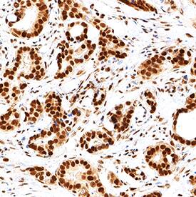
DDX1 was detected in immersion fixed paraffin-embedded sections of human breast cancer tissue using Rabbit Anti-Human/Mouse/Rat DDX1 Monoclonal Antibody (Catalog # MAB9615) at 3 µg/mL for 1 hour at room temperature followed by incubation with the Anti-Mouse IgG VisUCyte™ HRP Polymer Antibody (Catalog # VC001). Tissue was stained using DAB (brown) and counterstained with hematoxylin (blue). Specific staining was localized to cell nuclei. View our protocol for IHC Staining with VisUCyte HRP Polymer Detection Reagents.Applications for Human/Mouse/Rat DDX1 Antibody
Immunocytochemistry
Sample: Immersion fixed HeLa human cervical epithelial carcinoma cell line
Immunohistochemistry
Sample: Immersion fixed paraffin-embedded sections of human breast cancer tissue
Western Blot
Sample: Y‑79 human retinoblastoma cell line, A431 human epithelial carcinoma cell line, NIH‑3T3 mouse embryonic fibroblast cell line, Neuro‑2A mouse neuroblastoma cell line, and Rat‑2 rat embryonic fibroblast cell line
Formulation, Preparation, and Storage
Purification
Reconstitution
Formulation
Shipping
Stability & Storage
- 12 months from date of receipt, -20 to -70 °C as supplied.
- 1 month, 2 to 8 °C under sterile conditions after reconstitution.
- 6 months, -20 to -70 °C under sterile conditions after reconstitution.
Background: DDX1
DEAD (Asp-Glu-Ala-Asp) box helicase 1 (DDX1) is a putative RNA helicase gene. RNA helicases are involved in various cellular processes involving alteration of RNA secondary structure such as translation initiation, nuclear and mitochondrial splicing, and ribosome and spliceosome assembly. It has been proposed that DDX1 plays a role in RNA clearance at DNA double-strand breaks. It has also been reported as a coactivator of the NF-kappa-B promoter. DDX1 also acts as a positive transcriptional regulator of cyclin CCND2 expression. Human DDX1 has 97% homology to mouse and rat DDX1. Expression in embryos and testis suggest that DDX1 mediates cell growth and development. DDX1 has been proposed as a diagnostic marker for early recurrence in primary breast cancer.
Long Name
Alternate Names
Gene Symbol
UniProt
Additional DDX1 Products
Product Documents for Human/Mouse/Rat DDX1 Antibody
Product Specific Notices for Human/Mouse/Rat DDX1 Antibody
For research use only


