Human/Mouse/Rat GIT1 Antibody
R&D Systems, part of Bio-Techne | Catalog # MAB8508


Key Product Details
Species Reactivity
Applications
Label
Antibody Source
Product Specifications
Immunogen
Ser485-Asp636
Accession # Q9Y2X7
Specificity
Clonality
Host
Isotype
Scientific Data Images for Human/Mouse/Rat GIT1 Antibody
Detection of Human, Mouse, and Rat GIT1 by Western Blot.
Western blot shows lysates of HUVEC human umbilical vein endothelial cells, U2OS human osteosarcoma cell line, M1 mouse myeloid leukemia cell line, and Rat-2 rat embryonic fibroblast cell line. PVDF membrane was probed with 2 µg/mL of Mouse Anti-Human GIT1 Monoclonal Antibody (Catalog # MAB8508) followed by HRP-conjugated Anti-Mouse IgG Secondary Antibody (Catalog # HAF018). A specific band was detected for GIT1 at approximately 95 kDa (as indicated). This experiment was conducted under reducing conditions and using Immunoblot Buffer Group 1.GIT1 in Human Brain.
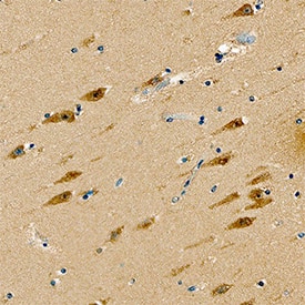
GIT1 was detected in immersion fixed paraffin-embedded sections of human brain (hippocampus) using Mouse Anti-Human GIT1 Monoclonal Antibody (Catalog # MAB8508) at 15 µg/mL overnight at 4 °C. Tissue was stained using the Anti-Mouse HRP-DAB Cell & Tissue Staining Kit (brown; Catalog # CTS002) and counterstained with hematoxylin (blue). Specific staining was localized to neurons. View our protocol for Chromogenic IHC Staining of Paraffin-embedded Tissue Sections.Detection of GIT1 in SH-SY5Y Human Cell line by Flow Cytometry.
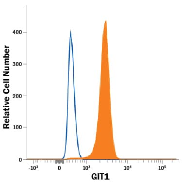
SH-SY5Y human neuroblastoma cell line was stained with Mouse Anti-Human GIT1 Monoclonal Antibody (Catalog # MAB8508, filled histogram) or isotype control antibody (Catalog # MAB0041, open histogram), followed by Allophycocyanin-conjugated Anti-Mouse IgG Secondary Antibody (Catalog # F0101B). To facilitate intracellular staining, cells were fixed with Flow Cytometry Fixation Buffer (Catalog # FC004) and permeabilized with Flow Cytometry Permeabilization/Wash Buffer I (Catalog # FC005).Applications for Human/Mouse/Rat GIT1 Antibody
CyTOF-ready
ELISA
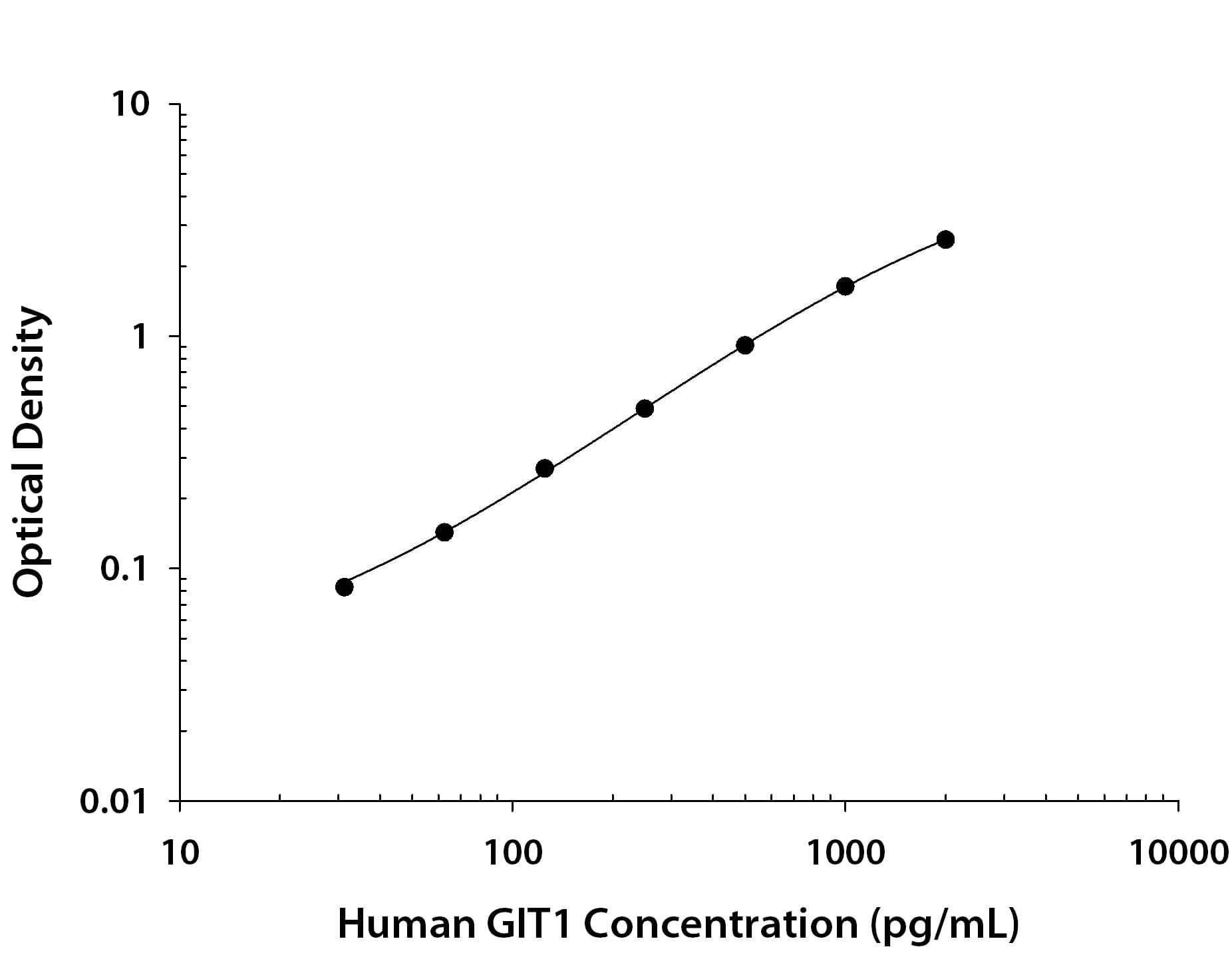
This antibody functions as an ELISA capture antibody when paired with Mouse Anti-Human GIT1 Monoclonal Antibody (Catalog # MAB85081). In sandwich immunoassays, this antibody is specific for human GIT1 when paired with the suggested detection antibody.
This product is intended for assay development on various assay platforms requiring antibody pairs. We recommend the Human GIT1 DuoSet ELISA Kit (Catalog # DY8485-05) for convenient development of a sandwich ELISA.
Immunohistochemistry
Sample: Immersion fixed paraffin-embedded sections of human brain (hippocampus)
Intracellular Staining by Flow Cytometry
Sample: SH‑SY5Y human neuroblastoma cell line fixed with Flow Cytometry Fixation Buffer (Catalog # FC004) and permeabilized with Flow Cytometry Permeabilization/Wash Buffer I (Catalog # FC005)
Western Blot
Sample: HUVEC human umbilical vein endothelial cells, U2OS human osteosarcoma cell line, M1 mouse myeloid leukemia cell line, and Rat‑2 rat embryonic fibroblast cell line
Formulation, Preparation, and Storage
Purification
Reconstitution
Formulation
Shipping
Stability & Storage
- 12 months from date of receipt, -20 to -70 °C as supplied.
- 1 month, 2 to 8 °C under sterile conditions after reconstitution.
- 6 months, -20 to -70 °C under sterile conditions after reconstitution.
Background: GIT1
Long Name
Alternate Names
Gene Symbol
UniProt
Additional GIT1 Products
Product Documents for Human/Mouse/Rat GIT1 Antibody
Product Specific Notices for Human/Mouse/Rat GIT1 Antibody
For research use only


