Human/Mouse/Rat Glutaredoxin 3/GLRX3 Antibody
R&D Systems, part of Bio-Techne | Catalog # AF7560

Key Product Details
Species Reactivity
Applications
Label
Antibody Source
Product Specifications
Immunogen
Asn126-Lys294
Accession # O76003
Specificity
Clonality
Host
Isotype
Scientific Data Images for Human/Mouse/Rat Glutaredoxin 3/GLRX3 Antibody
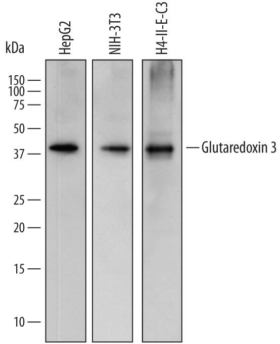
Detection of Human, Mouse, and Rat Glutaredoxin 3/GLRX3 by Western Blot.
Western blot shows lysates of HepG2 human hepatocellular carcinoma cell line, NIH-3T3 mouse embryonic fibroblast cell line, and H4-II-E-C3 rat hepatoma cell line. PVDF membrane was probed with 1 µg/mL of Sheep Anti-Human Glutaredoxin 3/GLRX3 Antigen Affinity-purified Polyclonal Antibody (Catalog # AF7560) followed by HRP-conjugated Anti-Sheep IgG Secondary Antibody (Catalog # HAF016). A specific band was detected for Glutaredoxin 3/GLRX3 at approximately 40 kDa (as indicated). This experiment was conducted under reducing conditions and using Immunoblot Buffer Group 1.Glutaredoxin 3/GLRX3 in Human Breast Cancer Tissue.
Glutaredoxin 3/GLRX3 was detected in immersion fixed paraffin-embedded sections of human breast cancer tissue using Sheep Anti-Human/Mouse/Rat Glutaredoxin 3/GLRX3 Antigen Affinity-purified Polyclonal Antibody (Catalog # AF7560) at 5 µg/mL overnight at 4 °C. Tissue was stained using the Anti-Sheep HRP-DAB Cell & Tissue Staining Kit (brown; Catalog # CTS019) and counterstained with hematoxylin (blue). Specific staining was localized to cytoplasm in ductal cells. View our protocol for Chromogenic IHC Staining of Paraffin-embedded Tissue Sections.Applications for Human/Mouse/Rat Glutaredoxin 3/GLRX3 Antibody
Immunohistochemistry
Sample: Immersion fixed paraffin-embedded sections of human breast cancer tissue
Western Blot
Sample: HepG2 human hepatocellular carcinoma cell line, NIH‑3T3 mouse embryonic fibroblast cell line, and H4‑II‑E‑C3 rat hepatoma cell line
Formulation, Preparation, and Storage
Purification
Reconstitution
Formulation
Shipping
Stability & Storage
- 12 months from date of receipt, -20 to -70 °C as supplied.
- 1 month, 2 to 8 °C under sterile conditions after reconstitution.
- 6 months, -20 to -70 °C under sterile conditions after reconstitution.
Background: Glutaredoxin 3/GLRX3
GLRX3 (Glutaredoxin-3; also PICOT, PKC-theta-interacting protein, and Thioredoxin-like protein 2/TXNL2) is a 37-40 kDa member of the multidomain subgroup, monothiol glutaredoxin group of glutaredoxin/GTX proteins. It has widespread expression, but is not found ubiquitously. Cells known to express cytosolic GLRX3 are principally epithelial in type, and include breast and adrenal epithelium, pancreatic exocrine gland epithelium, and proximal tubule renal epithelium. Cardiac muscle may also expresses GLRX3. The function of GLRX3 is not clear. By inference, it is suggested to be involved in cell proliferation, and has also been associated with both PKC-theta regulation and MLP/muscle LIM protein-mediated cardiac contractility. Human GLRX3 is 335 amino acids (aa) in length. It contains an N-terminal thioredoxin-like domain (aa 2-117) that is followed by two PICOT homology domains (aa 144-236 and 237-335). The thioredoxin domain does not possess a typical disulfide, and it is suggested that this domain does not demonstrate redox activity. The PICOT domains will interact with intracellular proteins. GLRX3 exists as both a monomer and homodimer, with the homodimer incorporating two Fe/S clusters into their PICOT domains. These clusters serve as redox sensors within the cell. Over aa 126-294, human GLRX3 shares 98% aa sequence identity with mouse GLRX3.
Alternate Names
Gene Symbol
UniProt
Additional Glutaredoxin 3/GLRX3 Products
Product Documents for Human/Mouse/Rat Glutaredoxin 3/GLRX3 Antibody
Product Specific Notices for Human/Mouse/Rat Glutaredoxin 3/GLRX3 Antibody
For research use only

