Human/Mouse/Rat Glypican 3 Antibody
R&D Systems, part of Bio-Techne | Catalog # MAB2119


Key Product Details
Species Reactivity
Validated:
Cited:
Applications
Validated:
Cited:
Label
Antibody Source
Product Specifications
Immunogen
Gln25-Val558
Accession # P51654.1
Specificity
Clonality
Host
Isotype
Scientific Data Images for Human/Mouse/Rat Glypican 3 Antibody
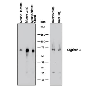
Detection of Mouse and Rat Glypican 3 by Western Blot.
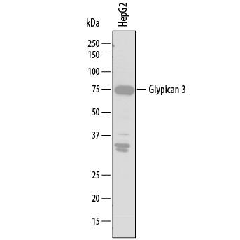
Western blot shows lysates of mouse placenta tissue, mouse lung tissue, mouse adrenal gland tissue, rat placenta tissue, and rat lung tissue. PVDF membrane was probed with 1 µg/mL of Mouse Anti-Human Glypican 3 Monoclonal Antibody (Catalog # MAB2119) followed by HRP-conjugated Anti-Mouse IgG Secondary Antibody (Catalog # HAF018). A specific band was detected for Glypican 3 at approximately 65-70 kDa (as indicated). This experiment was conducted under reducing conditions and using Immunoblot Buffer Group 1.Detection of Human Glypican 3 by Western Blot.
Western blot shows lysates of HepG2 human hepatocellular carcinoma cell line. PVDF membrane was probed with 2 µg/mL of Mouse Anti-Human Glypican 3 Monoclonal Antibody (Catalog # MAB2119) followed by HRP-conjugated Anti-Mouse IgG Secondary Antibody (Catalog # HAF007). A specific band was detected for Glypican 3 at approximately 75 kDa (as indicated). This experiment was conducted under reducing conditions and using Immunoblot Buffer Group 1.Glypican 3 in HepG2 Human Cell Line.
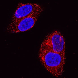
Glypican 3 was detected in immersion fixed HepG2 human hepatocellular carcinoma cell line using Mouse Anti-Human Glypican 3 Monoclonal Antibody (Catalog # MAB2119) at 3 µg/mL for 3 hours at room temperature. Cells were stained using the NorthernLights™ 557-conjugated Anti-Mouse IgG Secondary Antibody (red; Catalog # NL007) and counterstained with DAPI (blue). Specific staining was localized to cytoplasm and cell membranes. View our protocol for Fluorescent ICC Staining of Cells on Coverslips.Applications for Human/Mouse/Rat Glypican 3 Antibody
CyTOF-ready
Flow Cytometry
Sample: HepG2 human hepatocellular carcinoma cell line
Immunocytochemistry
Sample: Immersion fixed HepG2 human hepatocellular carcinoma cell line
Western Blot
Sample: HepG2 human hepatocellular carcinoma cell line, mouse placenta tissue, mouse lung tissue, mouse adrenal gland tissue, rat placenta tissue, and rat lung tissue
Reviewed Applications
Read 1 review rated 5 using MAB2119 in the following applications:
Formulation, Preparation, and Storage
Purification
Reconstitution
Formulation
Shipping
Stability & Storage
- 12 months from date of receipt, -20 to -70 °C as supplied.
- 1 month, 2 to 8 °C under sterile conditions after reconstitution.
- 6 months, -20 to -70 °C under sterile conditions after reconstitution.
Background: Glypican 3
Glypicans (GPC) are a family of heparan sulfate proteoglycans that are attached to the cell surface by a glycosylphosphatidylinositol (GPI) anchor. Six members of this family have been identified in mammals (GPC1-GPC6). All glypican core proteins contain an N-terminal signal peptide, a large globular cysteine-rich domain (CRD) with 14 invariant cysteine residues, a stalk-like region containing the heparan sulfate attachment sites, and a C-terminal GPI attachment site. While glypican proteins do not share strong amino acid sequence identity (they range from 17-63%), the conserved cysteine residues in their CRDs suggests similarity in their three‑dimensional structure (1, 2).
Mutations in GPC3 cause a rare disorder in humans, Simpson-Golabi-Behmel Syndrome, which is characterized by pre and postnatal overgrowth of multiple tissues and organs and an increased risk for developing embryonic tumors (3). These features are also present in the mouse knock-out of GPC3 indicating that GPC3 regulates cell survival and inhibits cell proliferation during development (4). Glypican 3 has been implicated in regulating many different signaling pathways including: IGF, FGF, BMP, and Wnt. An endoproteolytic processing of GPC3 by proprotein convertases is required for the modulation of Wnt signaling (5). Direct interaction with FGF-basic has been observed and is mediated by the heparan sulfate chains (6).
References
- Filmus, J. and S.B. Selleck (2001) J. Clinical Invest. 108:497.
- De Cat, B and G. David (2001) Seminars in Cell & Dev. Biol. 12:117.
- Pilia, G. et al. (1996) Nat. Genet. 12: 241.
- Cano-Gauci, D.F. et al. (1999) J. Cell Biol. 146: 255.
- De Cat, B. et al. (2003) J. Cell Biol. 163:625.
- Song, H.H. et al. (1997) J. Biol. Chem. 272:7574.
Alternate Names
Gene Symbol
UniProt
Additional Glypican 3 Products
Product Documents for Human/Mouse/Rat Glypican 3 Antibody
Product Specific Notices for Human/Mouse/Rat Glypican 3 Antibody
For research use only

