Human/Mouse/Rat Importin beta/KPNB1 Antibody
R&D Systems, part of Bio-Techne | Catalog # MAB8209

Key Product Details
Validated by
Biological Validation
Species Reactivity
Validated:
Human, Mouse, Rat
Cited:
Mouse
Applications
Validated:
Immunocytochemistry, Western Blot
Cited:
Western Blot
Label
Unconjugated
Antibody Source
Monoclonal Mouse IgG2B Clone # 845208
Product Specifications
Immunogen
E. coli-derived recombinant human Importin beta/KPNB1
Met1-Gly155
Accession # Q14974
Met1-Gly155
Accession # Q14974
Specificity
Detects human Importin beta/KPNB1 in ELISA and Western Blot. It detects mouse and rat Importin beta/KPNB1 in Western Blots.
Clonality
Monoclonal
Host
Mouse
Isotype
IgG2B
Scientific Data Images for Human/Mouse/Rat Importin beta/KPNB1 Antibody
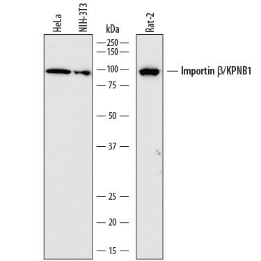
Detection of Human, Mouse, and Rat Importin beta/KPNB1 by Western Blot.
Western blot shows lysates of HeLa human cervical epithelial carcinoma cell line, NIH-3T3 mouse embryonic fibroblast cell line, and Rat-2 rat embryonic fibroblast cell line. PVDF membrane was probed with 1 µg/mL of Mouse Anti-Human Importin beta/KPNB1 Monoclonal Antibody (Catalog # MAB8209) followed by HRP-conjugated Anti-Mouse IgG Secondary Antibody (Catalog # HAF018). A specific band was detected for Importin beta/KPNB1 at approximately 97 kDa (as indicated). This experiment was conducted under reducing conditions and using Immunoblot Buffer Group 1.Importin beta/KPNB1 in HeLa Human Cell Line.
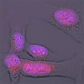
Importin beta/KPNB1 was detected in immersion fixed HeLa human cervical epithelial carcinoma cell line using Mouse Anti-Human Importin beta/KPNB1 Monoclonal Antibody (Catalog # MAB8209) at 25 µg/mL for 3 hours at room temperature. Cells were stained using the NorthernLights™ 557-conjugated Anti-Mouse IgG Secondary Antibody (red; Catalog # NL007) and counterstained with DAPI (blue). Specific staining was localized to nuclei. View our protocol for Fluorescent ICC Staining of Cells on Coverslips.Detection of Human Importin beta/KPNB1 by Western Blot
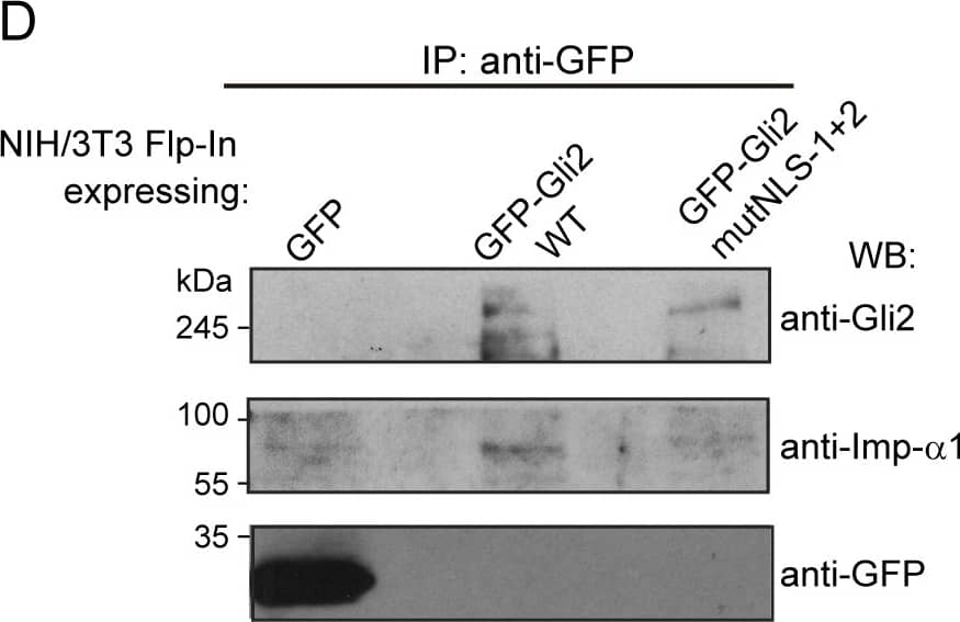
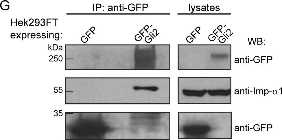
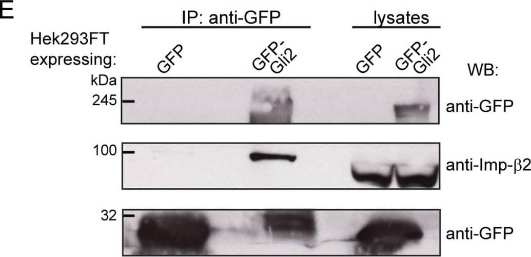
cNLSs are necessary for Gli2 nuclear import.(A) Sequences in Gli2_Mm predicted as cNLSs by cNLS-Mapper [40] using a cut-off of 5 with the corresponding scores (out of 10). Residues that were mutated for alanines in the mutNLS constructs are in bold. The mutated sequences are not predicted (n.p.) as cNLS. (B) Transduced NIH/3T3 cells expressing GFP-Gli2, wt or cNLS mutants (mutNLS-1, mutNLS-2 or mutNLS-1+2), were treated with SAG for 90 min or DMSO as control. Cells were stained for cilium (anti-Ac.Tub, red), GFP-Gli2 (anti-GFP, green) and nucleus (DAPI, blue). The same pictures are also shown with the channel corresponding to GFP-Gli2 in grey scale to help the visualization. Scale bar: 10 μm. (C) Quantification of nuclear Gli2 was performed as explained in Fig 1C. Black, solid line indicates comparison between control and Hh activated conditions for wtGli2, dotted line indicates comparison between wtGli2 under basal condition and mutNLS-1 or mutNLS-1+2 under basal or activated conditions and black, discontinued line indicates comparison between activated conditions for wtGl2 and mutNLS-2. *p<0.01, *** p<0.0001 (Kruskal-Wallis test). (B) and (C) are representative of four independent experiments and at least 70 cells were analysed for each condition. (D) WB detecting Imp-alpha 1 after precipitating GFP-Gli2 or GFP-Gli2-mutNLS-1+2 with GFP-Trap from NIH/3T3 Flp-In expressing at endogenous levels the constructs mentioned above. Membranes were cut at different levels so as to detect in the same samples the precipitated GFP-Gli2 (using an anti-Gli2 antibody) and GFP (left panels). Image collected and cropped by CiteAb from the following publication (https://pubmed.ncbi.nlm.nih.gov/27579771), licensed under a CC-BY license. Not internally tested by R&D Systems.Applications for Human/Mouse/Rat Importin beta/KPNB1 Antibody
Application
Recommended Usage
Immunocytochemistry
8-25 µg/mL
Sample: Immersion fixed HeLa human cervical epithelial carcinoma cell line
Sample: Immersion fixed HeLa human cervical epithelial carcinoma cell line
Western Blot
1 µg/mL
Sample: HeLa human cervical epithelial carcinoma cell line, NIH‑3T3 mouse embryonic fibroblast cell line, and Rat‑2 rat embryonic fibroblast cell line
Sample: HeLa human cervical epithelial carcinoma cell line, NIH‑3T3 mouse embryonic fibroblast cell line, and Rat‑2 rat embryonic fibroblast cell line
Reviewed Applications
Read 3 reviews rated 4.7 using MAB8209 in the following applications:
Formulation, Preparation, and Storage
Purification
Protein A or G purified from hybridoma culture supernatant
Reconstitution
Reconstitute at 0.5 mg/mL in sterile PBS. For liquid material, refer to CoA for concentration.
Formulation
Lyophilized from a 0.2 μm filtered solution in PBS with Trehalose. *Small pack size (SP) is supplied either lyophilized or as a 0.2 µm filtered solution in PBS.
Shipping
Lyophilized product is shipped at ambient temperature. Liquid small pack size (-SP) is shipped with polar packs. Upon receipt, store immediately at the temperature recommended below.
Stability & Storage
Use a manual defrost freezer and avoid repeated freeze-thaw cycles.
- 12 months from date of receipt, -20 to -70 °C as supplied.
- 1 month, 2 to 8 °C under sterile conditions after reconstitution.
- 6 months, -20 to -70 °C under sterile conditions after reconstitution.
Background: Importin beta/KPNB1
Long Name
Karyopherin beta 1
Alternate Names
IMB1, Impnb, Importin 1, IPOB, KPNB1, NTF97, PTAC97
Gene Symbol
KPNB1
UniProt
Additional Importin beta/KPNB1 Products
Product Documents for Human/Mouse/Rat Importin beta/KPNB1 Antibody
Product Specific Notices for Human/Mouse/Rat Importin beta/KPNB1 Antibody
For research use only
Loading...
Loading...
Loading...
Loading...
