Human/Mouse/Rat PD-ECGF/Thymidine Phosphorylase Antibody
R&D Systems, part of Bio-Techne | Catalog # MAB7568

Key Product Details
Species Reactivity
Applications
Label
Antibody Source
Product Specifications
Immunogen
Met1-Pro471
Accession # Q99N42
Specificity
Clonality
Host
Isotype
Scientific Data Images for Human/Mouse/Rat PD-ECGF/Thymidine Phosphorylase Antibody
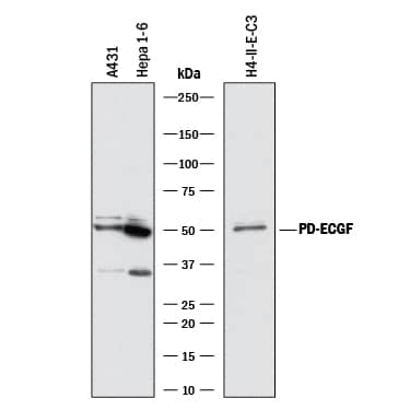
Detection of Human, Mouse, and Rat PD-ECGF/Thymidine Phosphorylase by Western Blot.
Western blot shows lysates of A431 human epithelial carcinoma cell line, Hepa 1-6 mouse hepatoma cell line, and H4-II-E-C3 rat hepatoma cell line. PVDF membrane was probed with 2 µg/mL of Rat Anti-Human/Mouse/Rat PD-ECGF/Thymidine Phosphorylase Monoclonal Antibody (Catalog # MAB7568) followed by HRP-conjugated Anti-Rat IgG Secondary Antibody (Catalog # HAF005). A specific band was detected for PD-ECGF/Thymidine Phosphorylase at approximately 49 kDa (as indicated). This experiment was conducted under reducing conditions and using Immunoblot Buffer Group 1.PD-ECGF/Thymidine Phosphorylase in Mouse Embryo.
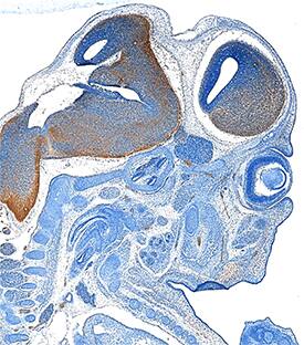
PD-ECGF/Thymidine Phosphorylase was detected in immersion fixed frozen sections of mouse embryo (15 d.p.c.) using Rat Anti-Human/Mouse/Rat PD-ECGF/Thymidine Phosphorylase Monoclonal Antibody (Catalog # MAB7568) at 25 µg/mL overnight at 4 °C. Tissue was stained using the Anti-Rat HRP-DAB Cell & Tissue Staining Kit (brown; Catalog # CTS017) and counterstained with hematoxylin (blue). Specific staining was localized to developing brain. View our protocol for Chromogenic IHC Staining of Frozen Tissue Sections.Applications for Human/Mouse/Rat PD-ECGF/Thymidine Phosphorylase Antibody
Immunohistochemistry
Sample: Immersion fixed frozen sections of mouse embryo (15 d.p.c.)
Western Blot
Sample: A431 human epithelial carcinoma cell line, Hepa 1‑6 mouse hepatoma cell line, and H4‑II‑E‑C3 rat hepatoma cell line
Formulation, Preparation, and Storage
Purification
Reconstitution
Formulation
Shipping
Stability & Storage
- 12 months from date of receipt, -20 to -70 °C as supplied.
- 1 month, 2 to 8 °C under sterile conditions after reconstitution.
- 6 months, -20 to -70 °C under sterile conditions after reconstitution.
Background: PD-ECGF/Thymidine Phosphorylase
TYMP (Thymidine phosphorylase/TP; also [PD]-ECGF/platelet-derived endothelial cell growth factor, gliostatin and TdRPase) is a 50-55 kDa member of the pyrimidine-nucleoside phosphorylase family of enzymes. TYMP/TP is both a cytosolic and secreted molecule that is expressed by a variety of cell types, including, macrophages, hepatocytes, endometrial gland epithelium, vascular smooth muscle and endothelial cells. It has also been found in select tumor cell types, and via an angiogenic activity, has been proposed to promote tumor growth. TYMP converts thymidine to thymine and 2-deoxy-ribose-1P, and it is the 2-deoxy-ribose component that is believed to promote endothelial cell migration (but not proliferation). This may be due to the fact that 2-deoxy-ribose induces reactive oxygen species, which drive the production of angiogenic factors, and that 2-deoxy-ribose also activates endothelial cell integrins. Mouse TYMP is 471 amino acids (aa) in length. It contains a phosphorylase domain (aa 96-348), followed by a C-terminal region (aa 374-448). Although TYMP circulates, there is no definitive signal sequence. TYMP is known to form homodimers. Full-length mouse TYMP (aa 1-471) shares 80% and 92% aa sequence identity with human and rat TYMP, respectively.
Long Name
Alternate Names
Gene Symbol
UniProt
Additional PD-ECGF/Thymidine Phosphorylase Products
Product Documents for Human/Mouse/Rat PD-ECGF/Thymidine Phosphorylase Antibody
Product Specific Notices for Human/Mouse/Rat PD-ECGF/Thymidine Phosphorylase Antibody
For research use only

