Human/Mouse/Rat PDK-1 Antibody
R&D Systems, part of Bio-Techne | Catalog # MAB864

Key Product Details
Species Reactivity
Validated:
Human, Mouse, Rat
Cited:
Mouse
Applications
Validated:
Immunocytochemistry, Simple Western, Western Blot
Cited:
Immunohistochemistry, Western Blot
Label
Unconjugated
Antibody Source
Monoclonal Mouse IgG2A Clone # 650308
Product Specifications
Immunogen
E. coli-derived recombinant human PDK-1
Asn411-Gln556
Accession # O15530
Asn411-Gln556
Accession # O15530
Specificity
Detects human, mouse, and rat PDK-1 in Western blots.
Clonality
Monoclonal
Host
Mouse
Isotype
IgG2A
Scientific Data Images for Human/Mouse/Rat PDK-1 Antibody
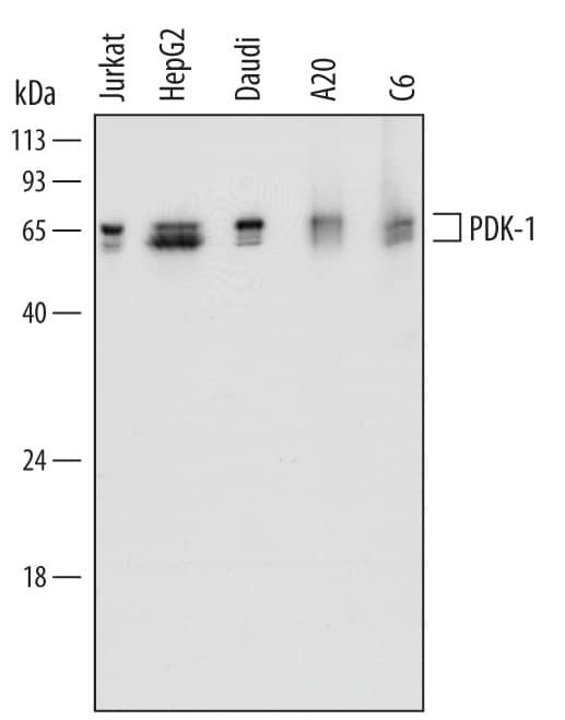
Detection of Human, Mouse, and Rat PDK‑1 by Western Blot.
Western blot shows lysates of Jurkat human acute T cell leukemia cell line, HepG2 human hepatocellular carcinoma cell line, Daudi human Burkitt's lymphoma cell line, A20 mouse B cell lymphoma cell line, and C6 rat glioma cell line. PVDF Membrane was probed with 1 µg/mL of Mouse Anti-Human/Mouse/Rat PDK-1 Monoclonal Antibody (Catalog # MAB864) followed by HRP-conjugated Anti-Mouse IgG Secondary Antibody (HAF007). Specific bands were detected for PDK-1 at approximately 58-68 kDa (as indicated). This experiment was conducted under reducing conditions and using Immunoblot Buffer Group 1.PDK‑1 in HeLa Human Cell Line.
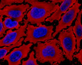
PDK-1 was detected in immersion fixed HeLa human cervical epithelial carcinoma cell line using Mouse Anti-Human/Mouse/Rat PDK-1 Monoclonal Antibody (Catalog # MAB864) at 10 µg/mL for 3 hours at room temperature. Cells were stained using the NorthernLights™ 557-conjugated Anti-Mouse IgG Secondary Antibody (yellow; NL007) and counterstained with DAPI (blue). Specific staining was localized to cytoplasm. View our protocol for Fluorescent ICC Staining of Cells on Coverslips.Detection of Human PDK-1 by Simple WesternTM.
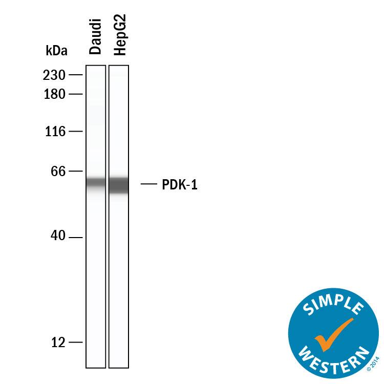
Simple Western shows lysates of Daudi human Burkitt's lymphoma cell line and HepG2 human hepatocellular carcinoma cell line, loaded at 0.5 mg/ml. A specific band was detected for PDK-1 at approximately 60 kDa (as indicated) using 20 µg/mL of Mouse Anti-Human/Mouse/Rat PDK-1 Monoclonal Antibody (Catalog # MAB864). This experiment was conducted under reducing conditions and using the 12-230 kDa separation system.Applications for Human/Mouse/Rat PDK-1 Antibody
Application
Recommended Usage
Immunocytochemistry
5-25 µg/mL
Sample: Immersion fixed HeLa human cervical epithelial carcinoma cell line
Sample: Immersion fixed HeLa human cervical epithelial carcinoma cell line
Simple Western
20 µg/mL
Sample: Daudi human Burkitt's lymphoma cell line and HepG2 human hepatocellular carcinoma cell line
Sample: Daudi human Burkitt's lymphoma cell line and HepG2 human hepatocellular carcinoma cell line
Western Blot
1 µg/mL
Sample: Jurkat human acute T cell leukemia cell line, HepG2 human hepatocellular carcinoma cell line, Daudi human Burkitt's lymphoma cell line, A20 mouse B cell lymphoma cell line, and C6 rat glioma cell line
Sample: Jurkat human acute T cell leukemia cell line, HepG2 human hepatocellular carcinoma cell line, Daudi human Burkitt's lymphoma cell line, A20 mouse B cell lymphoma cell line, and C6 rat glioma cell line
Formulation, Preparation, and Storage
Purification
Protein A or G purified from hybridoma culture supernatant
Reconstitution
Sterile PBS to a final concentration of 0.5 mg/mL. For liquid material, refer to CoA for concentration.
Formulation
Lyophilized from a 0.2 μm filtered solution in PBS with Trehalose. See Certificate of Analysis for details.
*Small pack size (-SP) is supplied either lyophilized or as a 0.2 µm filtered solution in PBS.
*Small pack size (-SP) is supplied either lyophilized or as a 0.2 µm filtered solution in PBS.
Shipping
Lyophilized product is shipped at ambient temperature. Liquid small pack size (-SP) is shipped with polar packs. Upon receipt, store immediately at the temperature recommended below.
Stability & Storage
Use a manual defrost freezer and avoid repeated freeze-thaw cycles.
- 12 months from date of receipt, -20 to -70 °C as supplied.
- 1 month, 2 to 8 °C under sterile conditions after reconstitution.
- 6 months, -20 to -70 °C under sterile conditions after reconstitution.
Background: PDK-1
Long Name
Phosphoinositide Dependent Kinase-1
Alternate Names
PDK1, PDPK1, PKB kinase
Gene Symbol
PDPK1
UniProt
Additional PDK-1 Products
Product Documents for Human/Mouse/Rat PDK-1 Antibody
Product Specific Notices for Human/Mouse/Rat PDK-1 Antibody
For research use only
Loading...
Loading...
Loading...
Loading...


