Human/Mouse/Rat Phospho-HSP27 (S78/S82) Antibody
R&D Systems, part of Bio-Techne | Catalog # AF2314

Key Product Details
Validated by
Biological Validation
Species Reactivity
Validated:
Human, Mouse, Rat
Cited:
Human
Applications
Validated:
Immunocytochemistry, Western Blot
Cited:
Flow Cytometry, Western Blot
Label
Unconjugated
Antibody Source
Polyclonal Rabbit IgG
Product Specifications
Immunogen
Phosphopeptide containing human HSP27 S78/S82 sites
Specificity
Detects human HSP27 when dually phosphorylated at S78/S82, and mouse and rat HSP27 phosphorylated at S86.
Clonality
Polyclonal
Host
Rabbit
Isotype
IgG
Scientific Data Images for Human/Mouse/Rat Phospho-HSP27 (S78/S82) Antibody
Detection of Human and Mouse Phospho-HSP27 (S78/S82) by Western Blot.
Western blot shows lysates of HeLa human cervical epithelial carcinoma cell line and C2C12 mouse myoblast cell line untreated (-) or treated (+) with 100 J/m2UV-C for 30 minutes. PVDF membrane was probed with 0.1 µg/mL of Rabbit Anti-Human/Mouse/Rat Phospho-HSP27 (S78/S82) Antigen Affinity-purified Polyclonal Antibody (Catalog # AF2314), followed by HRP-conjugated Anti-Rabbit IgG Secondary Antibody (Catalog # HAF008). A specific band was detected for Phospho-HSP27 (S78/S82) at approximately 27 kDa (as indicated). This experiment was conducted under reducing conditions and using Immunoblot Buffer Group 1.Phospho-HSP27 (S78/S82) in HeLa Human Cell Line.
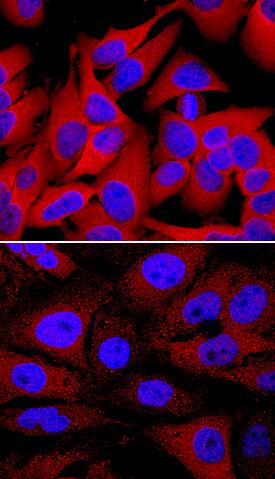
HSP27 phosphorylated at S78/S82 was detected in immersion fixed HeLa human cervical epithelial carcinoma cell line unstimulated (lower panel) or stimulated with 20 mJ/cm2ultraviolet radiation (upper panel) using Rabbit Anti-Human/Mouse/Rat Phospho-HSP27 (S78/S82) Antigen Affinity-purified Polyclonal Antibody (Catalog # AF2314) at 1 µg/mL for 3 hours at room temperature. Cells were stained using the NorthernLights™ 557-conjugated Anti-Rabbit IgG Secondary Antibody (red; Catalog # NL004) and counterstained with DAPI (blue). Specific staining was localized to cytoplasm and nuclei. View our protocol for Fluorescent ICC Staining of Cells on Coverslips.Detection of Human HSP27 by Western Blot
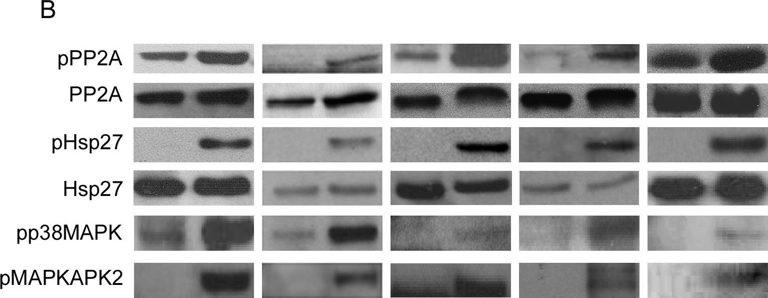
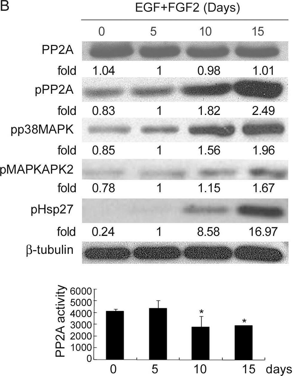
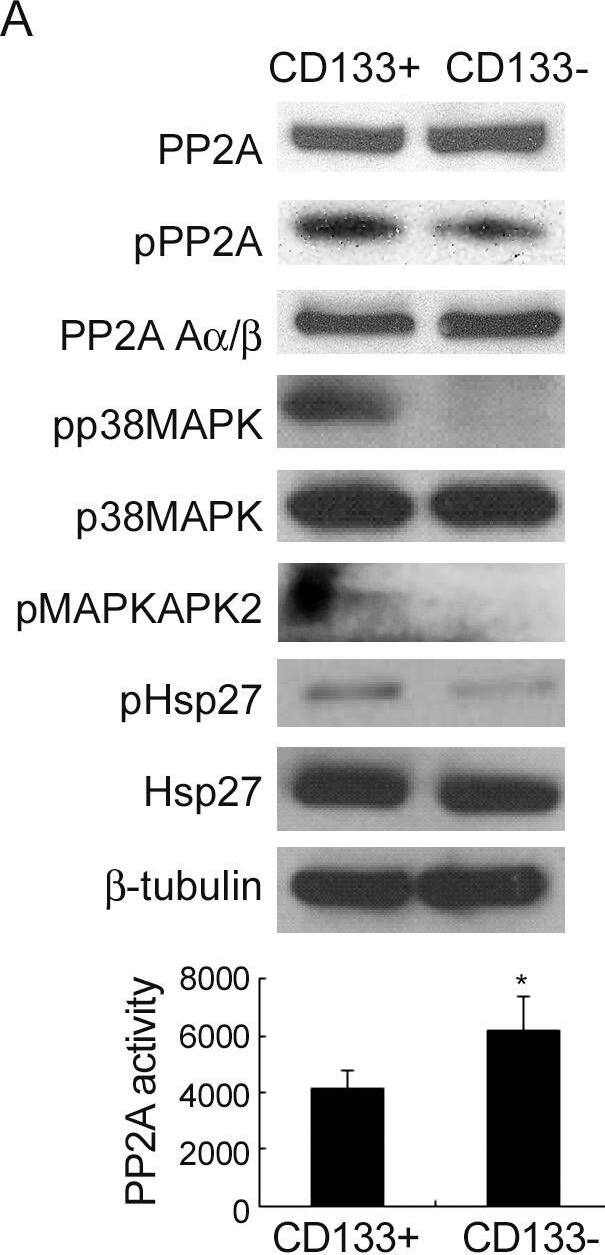
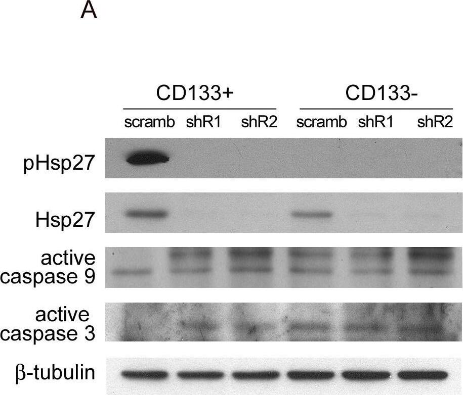
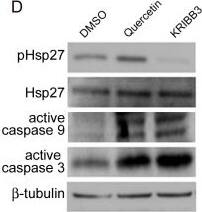
Enrichment of TICs decreases PP2A activity and increases Hsp27 activation in other solid tumors.CCS and HCW colorectal cancer cells, A549 lung cancer cells, HTB186 medulloblastoma cells and SAS, oral cancer cells were continually cultured under serum depletion in the presence of EGF (10 ng/mL) and FGF2 (10 ng/mL) to enrich TICs (E+F) or in serum-containing medium (CTR) as a control. (A) Immunoblots for pluripotent markers. (B) Immunoblots for phosphorylated PP2A (pPP2A), PP2A and Hsp27. (C) Immunoblots for caspase 9 and 3. (D) Schema demonstrates the antiapoptosis pathway of colorectal TICs in response to in vitro hypoxia and serum depletion. Image collected and cropped by CiteAb from the following publication (https://dx.plos.org/10.1371/journal.pone.0049605), licensed under a CC-BY license. Not internally tested by R&D Systems.Applications for Human/Mouse/Rat Phospho-HSP27 (S78/S82) Antibody
Application
Recommended Usage
Immunocytochemistry
5-15 µg/mL
Sample: Immersion fixed HeLa human cervical epithelial carcinoma cell line unstimulated or stimulated with 20 mJ/cm2 ultraviolet radiation
Sample: Immersion fixed HeLa human cervical epithelial carcinoma cell line unstimulated or stimulated with 20 mJ/cm2 ultraviolet radiation
Western Blot
0.1 µg/mL
Sample: UV-C-treated HeLa human cervical epithelial carcinoma cell line and C2C12 mouse myoblast cell line
Sample: UV-C-treated HeLa human cervical epithelial carcinoma cell line and C2C12 mouse myoblast cell line
Formulation, Preparation, and Storage
Purification
Antigen and protein A Affinity-purified
Reconstitution
Reconstitute at 0.2 mg/mL in sterile PBS. For liquid material, refer to CoA for concentration.
Formulation
Lyophilized from a 0.2 μm filtered solution in PBS with Trehalose. *Small pack size (SP) is supplied either lyophilized or as a 0.2 µm filtered solution in PBS.
Shipping
Lyophilized product is shipped at ambient temperature. Liquid small pack size (-SP) is shipped with polar packs. Upon receipt, store immediately at the temperature recommended below.
Stability & Storage
Use a manual defrost freezer and avoid repeated freeze-thaw cycles.
- 12 months from date of receipt, -20 to -70 °C as supplied.
- 1 month, 2 to 8 °C under sterile conditions after reconstitution.
- 6 months, -20 to -70 °C under sterile conditions after reconstitution.
Background: HSP27
References
- Gusev, N.B. et al. (2002) Biochemistry (Moscow) 67:511.
- Garrido, C. et al. (2001) Biochem. Biophys. Res. Commun. 286:433.
- Garrido, C. (2002) Cell Death Diffr. 9:483.
- Brvey, J-M. et al. (2000) Nat. Cell Biol. 2:645.
Long Name
Heat Shock Protein 27
Alternate Names
HSP25, HSPB1
Gene Symbol
HSPB1
Additional HSP27 Products
Product Documents for Human/Mouse/Rat Phospho-HSP27 (S78/S82) Antibody
Product Specific Notices for Human/Mouse/Rat Phospho-HSP27 (S78/S82) Antibody
For research use only
Loading...
Loading...
Loading...
Loading...
