Human/Mouse/Rat Phospho-RSK1 (S380) Antibody
R&D Systems, part of Bio-Techne | Catalog # MAB79671

Key Product Details
Validated by
Species Reactivity
Applications
Label
Antibody Source
Product Specifications
Immunogen
Accession # Q15418
Specificity
Clonality
Host
Isotype
Scientific Data Images for Human/Mouse/Rat Phospho-RSK1 (S380) Antibody
Detection of Human Phospho-RSK1 (S380) by Western Blot.
Western blot shows lysates of Jurkat human acute T cell leukemia cell line and HeLa human cervical epithelial carcinoma cell line untreated (-) or treated (+) with 200 nM PMA or PMA and Ionomycin for 20 minutes. PVDF membrane was probed with 0.1 µg/mL of Rabbit Anti-Human/Mouse/Rat Phospho-RSK1 (S380) Monoclonal Antibody (Catalog # MAB79671) followed by HRP-conjugated Anti-Rabbit IgG Secondary Antibody (Catalog # HAF008). A specific band was detected for Phospho-RSK1 (S380) at approximately 93 kDa (as indicated). This experiment was conducted under reducing conditions and using Immunoblot Buffer Group 1.RSK1 in HeLa Human Cell Line.
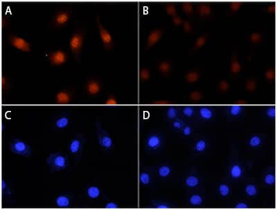
RSK1 was detected in immersion fixed HeLa human cervical epithelial carcinoma cell line, unstimulated (panels B and D) or stimulated with PMA (panels A and C), using Rabbit Anti-Human/Mouse/Rat Phospho-RSK1 (S380) Monoclonal Antibody (Catalog # MAB79671) at 25 µg/mL for 3 hours at room temperature. Cells were stained using the NorthernLights™ 557-conjugated Anti-Rabbit IgG Secondary Antibody (red, upper panels; Catalog # NL004) and counterstained with DAPI (blue, lower panels). Specific staining was localized to plasma membrances and cytoplasm. View our protocol for Fluorescent ICC Staining of Cells on Coverslips.Detection of Human Phospho-RSK1 (S380) by Simple WesternTM.
Simple Western lane view shows lysates of HeLa human cervical epithelial carcinoma cell line untreated (-) or treated (+) with 200 nM PMA for 20 minutes, and loaded at 0.2 mg/mL. A specific band was detected for Phospho-RSK1 (S380) at approximately 90 kDa (as indicated) using 1 μg/mL of Rabbit Anti-Human/Mouse/Rat Phospho-RSK1 (S380) Monoclonal Antibody (Catalog # MAB79671). This experiment was conducted under reducing conditions and using the 12-230 kDa separation system.Applications for Human/Mouse/Rat Phospho-RSK1 (S380) Antibody
Immunocytochemistry
Sample: Immersion fixed HeLa human cervical epithelial carcinoma cell line stimulated with PMA
Simple Western
Sample: HeLa human cervical epithelial carcinoma cell line treated with PMA
Western Blot
Sample: Jurkat human acute T cell leukemia cell line and HeLa human cervical epithelial carcinoma cell line treated with PMA or PMA and Ionomycin
Reviewed Applications
Read 1 review rated 5 using MAB79671 in the following applications:
Formulation, Preparation, and Storage
Purification
Reconstitution
Formulation
Shipping
Stability & Storage
- 12 months from date of receipt, -20 to -70 °C as supplied.
- 1 month, 2 to 8 °C under sterile conditions after reconstitution.
- 6 months, -20 to -70 °C under sterile conditions after reconstitution.
Background: RSK1
RSK1 (ribosomal S6 kinase 1), gene name RPS6KA1 (ribosomal protein S6 kinase alpha1), also called p90RSK1 (90 kDa RSK1) or MAPKAPK 1a (MAPK-activated protein kinase 1a), is a widely expressed member of the RSK family of growth factor-regulated serine/threonine kinases. RSK proteins contain two non-identical kinase catalytic domains, and mediate activation of mitogen-activated kinase (MAPK) cascades and stimulation of cell proliferation and differentiation. Autophosphorylation on S380 allows binding of PDPK1, leading to further phosphorylation and full activation of RSK1 kinase activity. Human, mouse and rat RSK1 share 98% aa sequence identity, and are identical within the region of the peptide immunogen.
Long Name
Alternate Names
Gene Symbol
UniProt
Additional RSK1 Products
Product Documents for Human/Mouse/Rat Phospho-RSK1 (S380) Antibody
Product Specific Notices for Human/Mouse/Rat Phospho-RSK1 (S380) Antibody
For research use only


