Human/Mouse/Rat PKC alpha Antibody
R&D Systems, part of Bio-Techne | Catalog # AF5340

Key Product Details
Species Reactivity
Validated:
Cited:
Applications
Validated:
Cited:
Label
Antibody Source
Product Specifications
Immunogen
Lys604-Val672
Accession # P17252
Specificity
Clonality
Host
Isotype
Scientific Data Images for Human/Mouse/Rat PKC alpha Antibody
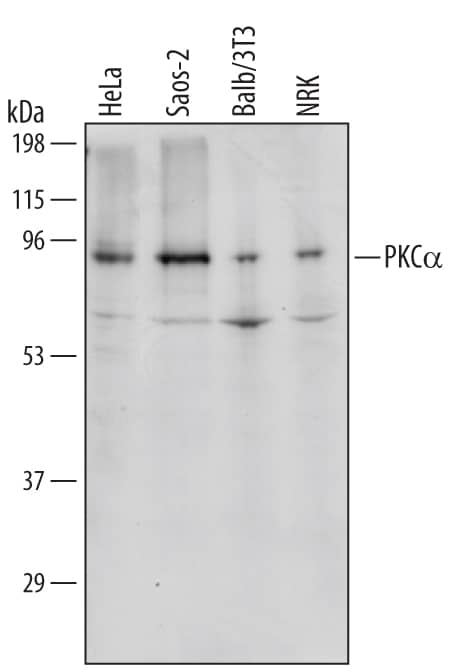
Detection of Human/Mouse/Rat PKC alpha by Western Blot.
Western blot shows lysates of HeLa human cervical epithelial carcinoma cell line, Saos-2 human osteosarcoma cell line, Balb/3T3 mouse embryonic fibroblast cell line, and NRK rat normal kidney cell line. PVDF membrane was probed with 1 µg/mL of Human/Mouse/Rat PKCa Antigen Affinity-purified Polyclonal Antibody (Catalog # AF5340) followed by HRP-conjugated Anti-Goat IgG Secondary Antibody (Catalog # HAF109). A specific band was detected for PKCa at approximately 90 kDa (as indicated). This experiment was conducted under reducing conditions and using Immunoblot Buffer Group 1.PKC alpha in Human Breast Cancer Tissue.
PKCa was detected in immersion fixed paraffin-embedded sections of human breast cancer tissue using Goat Anti-Human/Mouse/Rat PKCa Antigen Affinity-purified Polyclonal Antibody (Catalog # AF5340) at 10 µg/mL overnight at 4 °C. Tissue was stained using the Anti-Goat HRP-DAB Cell & Tissue Staining Kit (brown; Catalog # CTS008) and counterstained with hematoxylin (blue). Specific staining was localized to cancer cell cytoplasm. View our protocol for Chromogenic IHC Staining of Paraffin-embedded Tissue Sections.Detection of Human PKC alpha by Simple WesternTM.
Simple Western shows lysates of Saos-2 human osteosarcoma cell line, loaded at 0.5 mg/ml. A specific band was detected for PKC alpha at approximately 84 kDa (as indicated) using 10 µg/mL of Goat Anti-Human/Mouse/Rat PKC alpha Antigen Affinity-purified Polyclonal Antibody (Catalog # AF5340). This experiment was conducted under reducing conditions and using the 12-230kDa separation system.Applications for Human/Mouse/Rat PKC alpha Antibody
Immunohistochemistry
Sample: Immersion fixed paraffin-embedded sections of human breast cancer tissue
Simple Western
Sample: Saos-2 human osteosarcoma cell line
Western Blot
Sample: HeLa human cervical epithelial carcinoma cell line, Saos-2 human osteosarcoma cell line, Balb/3T3 mouse embryonic fibroblast cell line, and NRK rat normal kidney cells
Reviewed Applications
Read 3 reviews rated 5 using AF5340 in the following applications:
Formulation, Preparation, and Storage
Purification
Reconstitution
Formulation
*Small pack size (-SP) is supplied either lyophilized or as a 0.2 µm filtered solution in PBS.
Shipping
Stability & Storage
- 12 months from date of receipt, -20 to -70 °C as supplied.
- 1 month, 2 to 8 °C under sterile conditions after reconstitution.
- 6 months, -20 to -70 °C under sterile conditions after reconstitution.
Background: PKC alpha
PKC alpha (Protein kinase C alpha) is an 80 kDa member of the PKC subfamily, AGC Ser/Thr protein kinase family of enzymes. It is widely expressed, and serves multiple cell-specific functions. PKC alpha is activated by increased diacylglycerol and intracellular calcium, and translocates to several sites such as the Golgi, nucleus and plasma membrane. Human PKC alpha is 672 amino acids (aa) in length. It contains two zinc finger regions (aa 36‑151), a protein kinase domain (aa 339‑557), and an AGC kinase region (aa 598‑668). Phosphorylations on Thr497, Thr638 and Ser657 are necessary for kinase activity. Over aa 604‑672, human PKC alpha shows 100% aa identity to mouse PKC alpha.
Long Name
Alternate Names
Gene Symbol
UniProt
Additional PKC alpha Products
Product Documents for Human/Mouse/Rat PKC alpha Antibody
Product Specific Notices for Human/Mouse/Rat PKC alpha Antibody
For research use only


