Human/Mouse/Rat SHIP2 Antibody
R&D Systems, part of Bio-Techne | Catalog # AF5389

Key Product Details
Species Reactivity
Applications
Label
Antibody Source
Product Specifications
Immunogen
Ala1106-Lys1258
Accession # O15357
Specificity
Clonality
Host
Isotype
Scientific Data Images for Human/Mouse/Rat SHIP2 Antibody
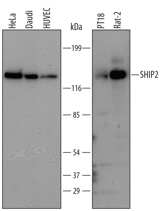
Detection of Human/Mouse/Rat SHIP2 by Western Blot.
Western blot shows lysates of HeLa human cervical epithelial carcinoma cell line, Daudi human Burkitt's lymphoma cell line, HUVEC human umbilical vein endothelial cells, PT18 mouse mast/basophil cell line, and Rat-2 rat embryonic fibroblast cell line. PVDF membrane was probed with 1 µg/mL of Sheep Anti-Human/Mouse/Rat SHIP2 Antigen Affinity-purified Polyclonal Antibody (Catalog # AF5389) followed by HRP-conjugated Anti-Sheep IgG Secondary Antibody (Catalog # HAF016). A specific band was detected for SHIP2 at approximately 140 kDa (as indicated). This experiment was conducted using Immunoblot Buffer Group 1.SHIP2 in PC‑3 Human Cell Line.
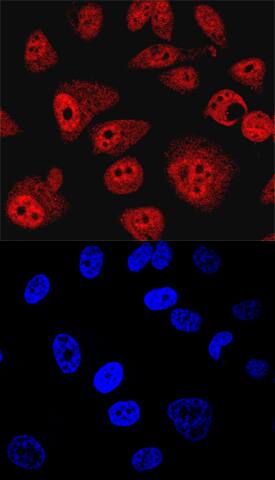
SHIP2 was detected in immersion fixed PC-3 human prostate cancer cell line using Sheep Anti-Human/Mouse/Rat SHIP2 Antigen Affinity-purified Polyclonal Antibody (Catalog # AF5389) at 10 µg/mL for 3 hours at room temperature. Cells were stained using the NorthernLights™ 557-conjugated Anti-Sheep IgG Secondary Antibody (red, upper panel; Catalog # NL010) and counterstained with DAPI (blue, lower panel). Specific staining was localized to nuclei and cytoplasm. View our protocol for Fluorescent ICC Staining of Cells on Coverslips. This application has not yet been tested in mouse or rat samples.SHIP2 in Human Breast Cancer Tissue.
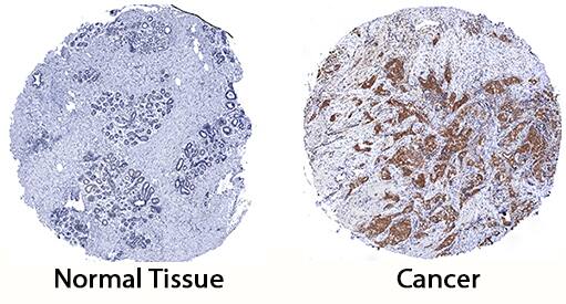
SHIP2 was detected in immersion fixed paraffin-embedded sections of human breast cancer tissue using Sheep Anti-Human/Mouse/Rat SHIP2 Antigen Affinity-purified Polyclonal Antibody (Catalog # AF5389) at 3 µg/mL for 1 hour at room temperature followed by incubation with the Anti-Goat IgG VisUCyte™ HRP Polymer Antibody (Catalog # VC004). Tissue was stained using DAB (brown) and counterstained with hematoxylin (blue). Specific staining was localized to cytoplasm in cancer cells. View our protocol for IHC Staining with VisUCyte HRP Polymer Detection Reagents.Applications for Human/Mouse/Rat SHIP2 Antibody
Immunocytochemistry
Sample: Immersion fixed PC‑3 human prostate cancer cell line
Immunohistochemistry
Sample: Immersion fixed paraffin-embedded sections of human breast cancer tissue
Western Blot
Sample: HeLa human cervical epithelial carcinoma cell line, Daudi human Burkitt's lymphoma cell line, HUVEC human umbilical vein endothelial cells, PT18 mouse mast/basophil cell line, and Rat-2 rat embryonic fibroblast cell line
Formulation, Preparation, and Storage
Purification
Reconstitution
Formulation
Shipping
Stability & Storage
- 12 months from date of receipt, -20 to -70 °C as supplied.
- 1 month, 2 to 8 °C under sterile conditions after reconstitution.
- 6 months, -20 to -70 °C under sterile conditions after reconstitution.
Background: SHIP2
SHIP2 (SH2 domain-containing inositol 5-phosphatase 2; also inositol polyphosphate phosphatase-like 1) is a member of the phosphatidylinositol/PtdIns 5-phosphatase family of enzymes. It is widely expressed and negatively regulates PI3 kinase pathways by hydrolyzing the 5-phosphate of PtdIns-3,4,5-triphosphate. Human SHIP2 is 1258 amino acids (aa) in length. It contains one SH2 domain (aa 21‑117), a Pro-rich region with an SH3 binding site (aa 935-1105), and a SAM (or sterile alpha-motif) domain that binds ARAP3 (aa 1196‑1258). SHIP2 is phosphorylated in response to growth factor activation on Tyr986, 1162 and 1358, and on Thr958, among other sites. Over aa 1106‑1258, human SHIP2 shares 95% aa identity with mouse SHIP2.
Long Name
Alternate Names
Gene Symbol
UniProt
Additional SHIP2 Products
Product Documents for Human/Mouse/Rat SHIP2 Antibody
Product Specific Notices for Human/Mouse/Rat SHIP2 Antibody
For research use only


