Human/Mouse/Rat Syntaxin 1A Antibody
R&D Systems, part of Bio-Techne | Catalog # AF7237

Key Product Details
Species Reactivity
Applications
Label
Antibody Source
Product Specifications
Immunogen
Met1-Leu165
Accession # Q16623
Specificity
Clonality
Host
Isotype
Scientific Data Images for Human/Mouse/Rat Syntaxin 1A Antibody
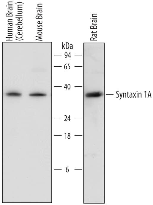
Detection of Human, Mouse, and Rat Syntaxin 1A by Western Blot.
Western blot shows lysates of human brain (cerebellum) tissue, mouse brain tissue, and rat brain tissue. PVDF membrane was probed with 0.2 µg/mL of Goat Anti-Human/Mouse/Rat Syntaxin 1A Antigen Affinity-purified Polyclonal Antibody (Catalog # AF7237) followed by HRP-conjugated Anti-Goat IgG Secondary Antibody (Catalog # HAF017). A specific band was detected for Syntaxin 1A at approximately 35 kDa (as indicated). This experiment was conducted under reducing conditions and using Immunoblot Buffer Group 1.Syntaxin 1A in SH-SY5Y Human Cell Line.
SH-SY5Y human neuroblastoma cells were cultured overnight in the presence of 1 mM Retinoic Acid (Catalog # 0695/50) prior to immersion fixation. Syntaxin 1A was detected using a Goat Anti-Human/Mouse/Rat Syntaxin 1A Antigen Affinity-purified Polyclonal Antibody (Catalog # AF7237). The cells were stained with the NorthernLights 557-conjugated Donkey Anti-Goat IgG Affinity-purified Secondary Antibody (red; Catalog # NL001). Actin filaments were stained with FITC-conjugated Phalloidin (green) and the cell nuclei were counter-stained with DAPI (blue). Syntaxin 1A immunoreactivity was localized to synaptic vesicles. View our protocol for Fluorescent ICC Staining of Cells on Coverslips.Applications for Human/Mouse/Rat Syntaxin 1A Antibody
Immunocytochemistry
Sample: Immersion fixed SH-SY5Y human neuroblastoma cell line
Western Blot
Sample: Human brain (cerebellum) tissue, mouse brain tissue, and rat brain tissue
Formulation, Preparation, and Storage
Purification
Reconstitution
Formulation
Shipping
Stability & Storage
- 12 months from date of receipt, -20 to -70 °C as supplied.
- 1 month, 2 to 8 °C under sterile conditions after reconstitution.
- 6 months, -20 to -70 °C under sterile conditions after reconstitution.
Background: Syntaxin 1A
STX1A (Syntaxin [Greek for 'organizing']1A; also HPC-1) is a 34-36 kDa member of the syntaxin family of proteins. It is a t-SNARE that is widely expressed in neurons, and is involved in the exocytosis of neurotransmitters at the presynaptic membrane. STX1A is transported intracellularly by microtubule-associated syntabulin, and its availability appears to be regulated through binding to LGI3. When released from LGI3, STX1A interacts with SNAP25 and VAMP2 to form the SNARE complex involved in exocytotic vesicle release. Human STX1A is a type IV single-pass transmembrane protein (very long cytoplasmic N-terminus) that is 288 amino acids (aa) in length. It contains a 265 aa N-terminal cytoplasmic domain that contains one coiled‑coil region (aa 68-109), a t-SNARE domain with a coiled‑coil region (aa 192-254), and a C-terminal transmembrane sequence (aa 266-286). There are three potential isoform variants. One shows a 34 aa substitution for aa 227‑288, while another termed STX1C is likely soluble, and contains a 25 aa substitution for the same aa sequence above encompassing aa 227‑288. A third variant shows a five aa substitution for aa 1-10. Over aa 1-165, human STX1A shares 99% aa sequence identity with mouse STX1A.
Alternate Names
Gene Symbol
UniProt
Additional Syntaxin 1A Products
Product Documents for Human/Mouse/Rat Syntaxin 1A Antibody
Product Specific Notices for Human/Mouse/Rat Syntaxin 1A Antibody
For research use only

